Searching for ease of use and predictability with resin cements
Indirect restorative materials have evolved rapidly over the past few years, giving clinicians a variety of options to better serve their patients. However, more choices in the substrate material often result in more steps in the cementation protocol, usually with specific and additional procedures that address the peculiarities of the chosen material. In addition, a cement chosen to increase retention of the indirect restoration comes at the cost of increased technique sensitivity and chair time to deliver the restoration.
Dental cement technology represents one of the major advancements in restorative dentistry. Older cements, such as zinc phosphate and zinc oxide eugenol, allowed for predictable retention of indirect restorations for more than a century, and various formulations are still used today.1 However, these materials may require protective measures in deeper preparations to avoid pulpal irritation, and they are susceptible to dissolution in water.2 Glass ionomer cements were invented in 1968, and, similar to earlier silicate-based products, they have the major benefit of releasing fluoride.1 However, while glass ionomer cement is easy to use, it is also water soluble. Even modern resin-modified varieties ideally require at least 15 minutes of protection from moisture contamination after placement.2
READ MORE | Give life to your restorations: Effective anterior composite contouring and finishing techniques
With advances and simplification of bonding protocols, resin cements have become more popular due to the high retention bond strengths that are achieved through micromechanical and chemical adhesion. Bonding is required for certain substrates, often due to fragility or minimal surface area available for adhesion to the tooth. This includes composite and ceramic inlays and onlays, as well as full-coverage restorations that may exhibit compromised angulation of the preparation or minimal height. Unlike most other cements, resin cements are insoluble in water and oral fluids.
Traditionally, resin cements required tooth preparation by etching, priming, curing, soft-tissue management to stop any bleeding, and isolating the tooth from moisture. These steps were time-consuming, and the tooth was often not optimally prepared due to technique sensitivity, such as excessive etching and desiccation of the dentin. Also, subgingival margins were often difficult to manage after exposure to the etchant and again after application of the adhesive.
Self-adhesive resin cements offer a simple solution to the traditional compromise between retentive bond strength and technique sensitivity. By combining the benefits of bonding with a cementation protocol that is easier than that used for traditional cements, self-adhesive resin cements provide superior retention with maximum simplicity. The self-adhesive feature means there is no need to apply etchant, primers, or adhesives to the prepared dental surfaces. This translates to greater predictability in preparations with subgingival margins, whereas etchants or bonding agents may cause bleeding.
Bisco's latest resin cement advances this concept with universal adhesion to all popular substrate materials. TheraCem is a dual-cured, calcium- and fluoride-releasing, self-adhesive resin cement indicated for luting crowns, bridges, inlays, onlays, and all types of posts. Delivering a strong bond to zirconia and most substrates, along with easy cleanup and high radiopacity, TheraCem offers reliable and durable cementation of most indirect restorations. Due to its unique chemistry, TheraCem achieves a high degree of conversion, which improves physical properties for added bond strength, without the need for refrigeration when not in use. This means peace of mind for clinicians and a product that is ready to use in every operatory.
Case No. 1
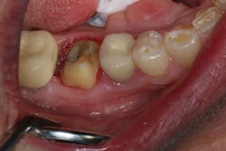
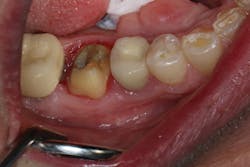
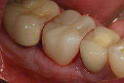
In the first case, a tooth with a subgingival margin preparation has been cleaned with an ultrasonic scaler and is gently dried in preparation for cementation. Note how the margin goes deeper subgingivally on the distolingual (figure 1). With TheraCem, a clean, prepped dentin or enamel surface is all that is needed to achieve excellent bond strengths, with the added benefit of sustained calcium and fluoride release. While TheraCem forms a strong bond to most substrates, including zirconia, a zirconia primer is still used prior to try-in to achieve optimal bond strength and prevent salivary contamination of the zirconia surface (figure 2).
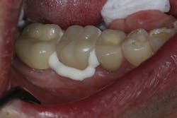
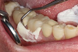
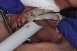
TheraCem exhibits minimal resistance to seating, but is not runny (figure 3). Cleanup is easy with hand instruments and floss (figure 4). For deeper subgingival margins, this cement is kind to the gingiva, although the margins should be thoroughly inspected to ensure complete removal of excess cement (figure 5).
Figure 1: Cleaned and prepared tooth
Figure 2: Z-Prime Plus single-component priming agent
Figure 3: TheraCem in use
Figure 4: TheraCem being cleaned up
Figure 5: Inspected margins
Case No. 2
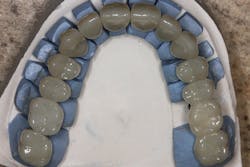
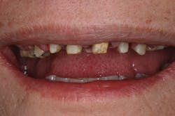
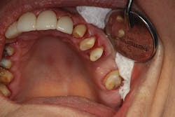
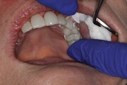
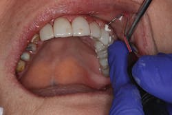
In the second case, a full arch of full-contour, anterior zirconia restorations are primed with zirconia primer and ready to be tried in (figure 6). Due to the patient's short clinical crowns, TheraCem was chosen to provide maximum retention of the zirconia restorations. In this case, attrition due to bruxism had reduced the height of the patient's teeth, and almost no vertical reduction was performed from first premolar to first premolar (figure 7). After try-in, the prostheses were rinsed and dried, and the front four crowns were delivered with TheraCem (figure 8). After preliminary cleanup of the cement, the upper left teeth were rinsed and gently dried (figure 9). The upper left restorations were then delivered with TheraCem (figure 10). This was followed up with another preliminary cleanup of the cement (figure 11). A retracted view shows the final restorations after a thorough cleanup (figure 12).
READ MORE | What restorative digital dentistry strategy should I embrace?
In both of these cases, where subgingival margins were present around each preparation, TheraCem was simple to use, easy to clean up, and did not require etching and priming each tooth prior to cementation. This resulted in a measurable decrease in chair time and frustration for both clinician and patient-especially in the second case, which was delivered in less than 30 minutes after administration of local anesthesia.
Figure 6: Restorations ready for try-in
Figure 7: Patient's teeth before restoration
Figure 8: Delivery of front crowns
Figure 9: Rinsed and dried upper left teeth
Figure 10: Delivery of upper left restorations
Figure 11: Preliminary cement cleanup
Figure 12: Final restorations after cleanup
References
1. Schulein TM. Significant events in the history of operative dentistry. J Hist Dent. 2005;53(2):63-72.
2. Weiner R. Liners, bases, and cements: material selection and clinical applications. Dentistry Today. 2005;24(6):66-72.
Joseph Kim, DDS, JD, received his dental training from the University of Michigan. He has maintained a general practice focused on sedation dentistry and full-arch implant therapy in the Chicago suburbs since 2002. Dr. Kim is excited to serve as a consultant to Bisco, Inc.
