Benefits of the diagnostic: Digitally designed or waxed
Sandy Cook, CDT, AAACD
The diagnostic wax-up is a useful tool to enhance treatment planning and provide a template for predictable restorations. The diagnostic is one of the first steps you should take when starting an esthetic case. By beginning with the end result in mind, a case that has a clear visual of the final product will go much more smoothly. In this article, I will discuss the merits of using a diagnostic wax-up—whether fabricated in wax or printed digitally—and how it can help optimize clinical and laboratory procedures to achieve the desired result.
In my experience, the diagnostic wax-up is a separate procedure that is usually performed by a technician in a laboratory. However, it can also be performed in the dental office if a more hands-on approach is desired while designing your case. Wax is applied to a model of the patient’s teeth, and stone is removed where necessary to create the proposed ideal smile design, as well as a template for the restorations. This is done in advance—before proceeding with the case—and it is often shown to the patient before treatment is accepted.
The term mock-up is often used interchangeably with wax-up. However, it is my understanding that a mock-up refers to a different kind of temporary diagnostic procedure—one that is performed directly in the patient’s mouth with composite. It can also be designed digitally, milled, and fitted over the patient’s existing teeth for a quick, temporary visual of the proposed treatment. This is like a preview smile for instant communication purposes and is usually removed shortly thereafter. A mock-up can be utilized before or after the actual diagnostic is made, but usually only when minor changes are indicated.
The diagnostic wax-up takes the mock-up a step further by refining and idealizing the outcome of the proposed restorations; it is used as a template for the completion of the case. For this article, I will focus mainly on the benefits of the diagnostic procedure.
A preview of the final proposed restorations
The diagnostic wax-up is undoubtedly one of the most important tools used to communicate between the clinician, patient, and dental laboratory technician. During this phase, complete pre-op dental records and photos of the patient are crucial and need to be sent with the case to the lab. In addition, any desires or concerns that the patient may have regarding his or her teeth must also be included in the case information. Sometimes the patient has an idea of what he or she likes and can offer to include pictures of magazines—or even old photos—of the desired result.
All of these records are used in the laboratory to reconstruct the patient’s presenting condition as accurately as possible. This also helps the technician visualize the patient and maybe get to know that person a little better, even if they’ve never met. I like to personalize each case if I can, because each patient is different. We must remember that the patients are the ones who have to look at themselves in the mirror each day and see the final results.
Once all of the records are gathered and the case is assembled, you have a clearer picture of how to proceed with the design. A wax or digital design of the case is then created. Every alteration is worked out in this step, including incisal length, tooth shape, function, occlusion, and any proposed tissue recontouring. A proposed model of the prep design and tissue recontouring is sometimes necessary and may even help to determine any unforeseen periodontal or implant concerns.
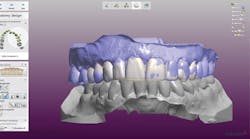
This same step can be seen in the digital design process, and screenshots during the design phase can be emailed to the clinician for discussion if needed (figure 1). This process will help the clinician and lab determine if certain restorations can be minimally prepped veneers or if full-coverage crowns, bridges, or implants would be implicated. The technician should be able to use this process to communicate any concerns that may arise during the fabrication and contact the doctor at that time to discuss any changes that need to be addressed before proceeding.
Figure 1: Screenshot of digital design in progress
After completing the design, a final model is made and sent to the doctor, along with a model of the original teeth. The changes in the diagnostic are often dramatic ones and will serve as the foundation for the treatment plan. These photos show a wax diagnostic both before and after in addition to a digitally printed diagnostic before and after (figures 2–5).
Figure 2: Pre-op before wax
Case presentation and acceptance
Each case is unique. Once the diagnostic is fabricated and returned to the doctor’s office, it can be very useful in presenting the patient with an idea of what the final restorations will look like. It can create a certain excitement and willingness for the patient to be more open to treatment, as well as a desire to move forward. It may be the first time the patient can actually see the possibilities for his or her new smile.
Patient education tool and peace of mind for you
In addition to serving as a communication tool for esthetic purposes, the diagnostic wax-up can also be used as a visual aid to help the patient understand his or her unique situation. Sometimes reality is a little different from the patient’s expectations. Having a clear representation of the desired outcome right out in front helps avoid unnecessary chairside disasters. Any esthetic likes, dislikes, or changes can be noted and discussed.
Figure 3: Diagnostic in wax showing suggested tissue recontouring
At this point, changes are very easy to do. Adding or removing wax on a model or altering a digital design is much easier to do than if the case were designed in porcelain without a diagnostic. Returning porcelain for changes is another matter. We don’t want to go there if we can help it. A good diagnostic wax-up can help avoid a scenario which might otherwise result in costly remakes and frustration for everyone. Diagnostics are designed to be a representation of what is actually possible for the dentition of each individual case.
Figure 4: Pre-op before digital design
Guide for determining preparation design
During the fabrication of the diagnostic, adding and removing proposed tooth structure in certain areas helps determine the ideal prep design for the case. As a technician, it is much easier to remove stone, add wax, or move things digitally. I try to be cognizant of the patient in the chair being prepped as I work on the diagnostic, as well as the doctor who is removing the tooth structure, so I am careful to reduce only as much as needed. The more clearly I can indicate in the design where to reduce porcelain, where to add porcelain, and where to reduce tissue, the easier it will be for the clinician and the patient. Typically, I will indicate any suggested tissue recontouring in the final diagnostic model and mark it so it can be duplicated during preparation. If it is more than a very small amount, a surgical stent is made and surgery can be performed if necessary. A second putty matrix (Sil-Tech, Ivoclar Vivadent) of the definitive wax-up can be used in the preparation process by slicing it in various ways to ensure that there is sufficient space for the proposed restorations (figure 6). Additional matrices can also be made at chairside.
Figure 5: 3-D digitally printed diagnostic
Blueprint for fabricating temporaries
The provisionals are created using a putty matrix made from the final diagnostic wax-up or 3-D–printed digital model. Sometimes, a light-body impression material is used as a wash inside the matrix to achieve a more detailed version of the model and to help incorporate these details into the temporaries. At this point, changes can be made in the temporaries, if needed, and a final impression taken. Sometimes, this process can take a few attempts to make sure the patient is satisfied with the result, but our goal is to help make this process faster and easier, with the final temporaries looking very much like the diagnostic model.
Figure 6: Sil-tech stents of final diagnostic with and without wash
The importance of the diagnostic wax-up and its merits cannot be overstated. Fabricating an esthetic case with a proper diagnostic wax-up is like having a solid foundation to build on for the duration of the treatment. Without a diagnostic, esthetic cases can be hit or miss. We all want every case to be a hit. It is much easier to incorporate the diagnostic process on a regular basis for more predictable and desirable results.
Sandy Cook, CDT, AAACD, is a ceramist at Microdental Northwest Laboratory in Kennewick, Washington. During the last 34 years, she has worked with leaders in cosmetic dentistry, including Matt Roberts at CMR Dental Lab in Idaho, where she worked for 21 years before moving to Washington. She is an accredited member of the American Academy of Cosmetic Dentistry. Contact her by email at [email protected].


