Smile makeover: A minimally invasive recipe for esthetic success
According to a recent survey, one-third of American adults are unhappy with their smiles, and 36% believe they would have better social lives with improved smiles. Growing up with social media has led to an increased dissatisfaction among young adults regarding their smiles. Seventy percent of those polled are self-conscious about their smiles, 57% cover their smiles when they laugh, and 48% have untagged themselves from social media posts because they did not like their smiles in the photos.1
Another study found that the biggest dating turnoff among men and women was “bad teeth.” According to the study, 77% of women think crooked teeth are worse than a receding hairline, and 78% of Americans perceive adults with crooked teeth to be unsuccessful in their business profession.2 Surprisingly, even though so many Americans are self-conscious about their teeth, the willingness to go to the dentist and get them fixed is not as popular as you may think.
When these same people with low smile confidence were polled, 30% said they would rather watch the same movie three times in a row than go to the dentist, and 25% said they would rather babysit an infant for three hours.3 When asked why people did not go to the dentist or had dental phobia, the invasive or intrusive nature of dentistry was cited as being in the top three.4
Read more:
- The treatment planning dilemma: To restore or replace the natural dentition
- The biomimetically driven restorative practice
To enable patients who either have dental phobia or “do not like going to the dentist” to achieve confident smiles, suggested treatment modalities include minimally invasive dentistry, short treatment times, fewer required dental visits, decreased postoperative pain, and a comprehensive understanding between dentist and patient.5 In this article, we will present a minimally invasive interdisciplinary case study that employed these techniques to achieve an esthetic smile makeover in a young adult with dental fear.
Case presentation

Visit 1
At the first appointment, a full-mouth series of radiographs, photographs, and dental records were obtained. Impressions were taken for a diagnostic wax-up, and the patient assisted in selecting the shape of her teeth (figure 3). I recommended making a preparation guide from the wax-up as this greatly assists the clinician while the teeth are prepared (figure 4). The preparation guide assures the clinician that adequate tooth structure is removed.
Visit 2
Because the size of the teeth is one of the most noticeable factors in a smile, it is important that the teeth look proportional and have an ideal width-to-height ratio.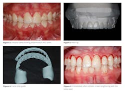
The patient was sent to the periodontal specialist, who used a 9.3-micron CO2 Solea laser (Convergent Dental) in a minimally invasive closed-flap esthetic crown lengthening procedure. No sutures were needed, and total chair time took less than 30 minutes to achieve the desired gingival height and shape (figure 5).
One study suggests that if surgical time can be cut down by just 10 minutes, moderate to severe postoperative pain can be decreased by as much as 40%.6 Other studies have suggested that laser therapy can expedite the healing of hard and soft tissue by stimulating growth factor release, which may cut down on the amount of time needed to heal after surgery.7 In this case, four weeks of gingival healing was needed prior to initiation of cosmetic restoration.
Visit 3
After the teeth were minimally prepared using a preparation guide, a hydrophilic polyvinyl siloxane (PVS) impression material, Splash Max (DenMat), was used. Additionally, an occlusal record, Artex facebow, stick bite, preparation shades, final impressions, bite registration, central length measurements, and photos were taken. The teeth were cleaned with chlorhexidine for 15 seconds and air-dried. A coat of Gluma desensitizer (Kulzer) was placed on the teeth to minimize any postoperative sensitivity.
From the wax-up, a PVS putty was made to help fabricate the provisional more quickly. Provisionals were made with Luxatemp Ultra (DMG) bleach light shade,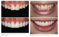
An impression of the provisional was taken and sent to the laboratory, along with a detailed written prescription of the materials and shades for the final porcelain restorations. Instructions to the lab were to follow the provisional as a guide for the size, shape, and contour.
Note: A well-done provisional is critical in a smile design case because the provisional serves as an important communication tool once the patient approves the smile design and gives the dental lab a blueprint to follow. The esthetics, phonetics, and bite can be inspected and changed before the final restoration is made by the dental laboratory.
Visit 4
After local anesthesia was administered and an OptraGate (Ivoclar Vivadent) rubber dam was placed, the provisional was removed and the veneers were tried in for verification of fit and esthetics. A neutral shade try-in gel (Ivoclar Vivadent) was used, and the patient approved the esthetics, color, and shape of the veneers. Then the patient signed off on the consent form for cementation of the veneers.
Note: A consent form for cementation is a very important step in the verification process, because once the veneers are cemented, changing them can be laborious and costly.
Note: If there is bleeding around the gums, apply Superoxyl (Sultan) along the gum margins and rinse.
The teeth were cleaned with hydrogen peroxide and chlorhexidine, Consepsis (Ultradent Product), in preparation for bonding. The teeth were then etched with 35% phosphoric acid (Ultradent) for 20 seconds, rinsed, and lightly dried. A coat of Gluma desensitizer was placed on the teeth to minimize any postoperative sensitivity and the teeth were air-dried. A single coat of Adhese Universal (Ivoclar Vivadent) was applied on the prepped teeth, agitated for 20 seconds, and then air-thinned and light-cured. After the excess solvent had evaporated, the teeth were cured with the Optilux 501 light (Demetron/Kerr) for 10 seconds per tooth. Ten feldspathic porcelain veneers with the Variolink Esthetic (Ivoclar Vivadent) were cemented and light-cured using neutral shade cement. The excess luting cement was cleaned off and all the veneers were tacked into place at the gingival crest for five seconds.
Note: Be sure the curing light is directed apically so there is no premature interproximal curing.
After all the veneers were tacked into place, a serrated saw blade was used interproximally and light-cured for five seconds per tooth. The teeth were carefully flossed, and excess cement was removed. All the porcelain veneers were completely cured for 40 seconds per surface and cleaned. Next, a thin coat of liquid strip (Ivoclar Vivadent) was applied on the margins of all the veneers and light-cured. Finally, the bite was checked for proper occlusion and polished.
Note: Liquid strip is a glycerin gel that prevents the oxygen-inhibited layer of composites with composite or ceramic restorations.
Visit 5
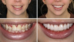
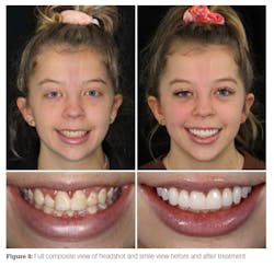
One week later, the patient returned to the office for a post-op check, and any necessary bite adjustments and/or esthetic recontouring were performed. Final photographs were taken and before-and-after photos were evaluated (figures 6–8).
In conclusion
This was an example of a patient who was extremely self-conscious about her smile but avoided treatment because of her dental fear. She avoided smiling, put her hand over her smile while laughing, and avoided being in pictures. With minimally invasive dentistry, planning, and great team communication, we were able to decrease healing times, chair time, and the number of required appointments. According to a written testimonial from the patient, she “is not embarrassed anymore about smiling and taking pictures. I am more confident in social settings and love hearing from my friends and family about how natural and beautiful my smile looks.”
Final note: In interdisciplinary cases such as this one, it is imperative that the clinician have extensive knowledge in the areas of smile design and case preparation. It is important to work as a team with your specialist and the patient to ensure a successful clinical outcome.
Editor's note: This article appeared in the April 2022 print edition of Dental Economics magazine. Dentists in North America are eligible for a complimentary print subscription. Sign up here.
References
- Study conducted by OnePoll with a sample of 2,000 Americans. OnePoll. 2018.
- Study shows that one-third of American adults are unhappy with their smile. American Association of Orthodontists. Study conducted by Wakefield Research. 2012.
- SWNS. More than half of Americans feel insecure about their teeth. New York Post. January 24, 2019. https://nypost.com/2019/01/24/more-than-half-of-americans-feel-insecure-about-their-teeth/
- Beaton L, Freeman R, Humphris G. Why are people afraid of the dentist? Observations and explanations. Med Princ Pract. 2014;23(4):295-301. doi:10.1159/000357223
- Slovin M. Managing the anxious and phobic dental patient. N Y State Dent J. 1997;63(7):36-40.
- Griffin TJ, Cheung WS, Zavras AI, Damoulis PD. Postoperative complications following gingival augmentation procedures. J Periodontol. 2006;77(12): 2070-2079. doi:1902/jop.2006.050296
- Arany PR, Cho A, Hunt TD, et al. Photoactivation of endogenous latent transforming growth factor–β1 directs dental stem cell differentiation for regeneration. Sci Transl Med. 2014;6(238):238ra69. doi:1126/scitranslmed.3008234
About the Author
Scott Froum, DDS
Scott Froum, DDS, a graduate of the State University of New York, Stony Brook School of Dental Medicine (SUNY), is a periodontist in private practice in New York City. He is the editorial director of Perio-Implant Advisory and serves on the Dental Economics advisory board. Dr. Froum is a volunteer professor in the postgraduate periodontal program at SUNY, is a trained naturopath, and is the scientific director of Meraki Integrative Functional Wellness Center. Contact him at drscottfroum.com.
Read Dr. Froum's DE Editorial Advisory Board profile here.

Michael J. Wei, DDS, FIADFE
Michael J. Wei, DDS, FIADFE, is a graduate of the New York University College of Dentistry. He received dual certificates in Advanced Education in General Dentistry (AEGD) at Columbia University School of Oral and Dental Surgery and completed a fellowship at New York Presbyterian Hospital. He currently is a clinical associate professor at New York University College of Dentistry. He maintains a private practice in Manhattan, New York, specializing in cosmetic dentistry and implant dentistry. Contact him through his website at mymanhattancosmeticdentist.com/ or (212) 982-4080.
Updated March 7, 2022