Incipient enamel caries: Etiology, detection, and treatment
The process of caries occurs due to the acid release from plaque bacteria. The extended ecological plaque hypothesis states that in conditions of health with infrequent glucose consumption and the presence of salivary buffers, commensal bacterial (nonmutans streptococci and actinomyces) proliferate. These bacteria neutralize acid through ammonia production and lactic acid metabolism.
With moderate and frequent exposure to acid, however, these bacteria colonies select more acidogenic strains that expel more protons in response to glucose metabolism. After extended periods at low pH, S. mutans and lactobacillus proliferation occurs. These bacteria are more acidogenic, meaning they produce a greater amount of lactic acid. Additionally, S. mutans produces extracellular polysaccharides such as glucans, which aid in adherence to enamel surfaces.1
The start of the caries process begins when hydrogen ions (acid) produced by bacteria in the biofilm covering teeth dissolve individual crystals of hydroxyapatite at a faster rate than they can be replenished. Enamel microstructure is composed of 70-170 nm long hydroxyapatite crystals arranged into 5-micron diameter enamel rods transversing lengthwise in an external-pulpal direction.2,3
The interior of the rods is referred to as prismatic enamel, which is surrounded by interprismatic (inter-rod) enamel oriented in a different direction. In between the two layers is a rod sheath composed of mostly noncollagenous proteins.3 Dissolution begins at the interface between prismatic and interprismatic dentin, leading to channels running alongside the length of the enamel rods through which hydrogen ions travel.
Dissolution of prismatic enamel on the outer 10-30 microns of the surface of the tooth is prevented by the presence of acid-resistant fluorapatite and demineralization-inhibiting macromolecules, such as proline-rich proteins and statherin, which are too large to penetrate deeper in enamel. The hydrogen ions that penetrate deep into enamel eventually dissolve prismatic enamel, creating the body of a subsurface “white spot lesion.”
Hydrogen ions can continue to travel through enamel until they reach the dentinoenamel junction. At that point, the dentin responds by odontoblasts-inducing mineralization in dentinal tubules, known as sclerotic dentin.4
Once sufficiently weakened, the external surface of the enamel may become cavitated through external mechanical damage from mastication, tooth contact, microtrauma, or even iatrogenic damage from dental probing.5 The enamel surface may then become infected with bacteria penetrating the prismatic enamel and eventually reaching the dentinoenamel junction (DEJ). Bacteria begin to infect dentinal tubules and eventually degrade intertubular dentin.4 Therefore, cavitation of enamel is a turning point in which a carious lesion would no longer be considered an incipient lesion.
Identification of incipient enamel lesions
Smooth surface incipient lesions are visualized as white spot lesions (WSLs). The subsurface porosity caused by dissolution of prismatic enamel changes the index of refraction of this enamel, creating the opacity of WSLs.
Visualization of WSLs may be improved by commercially available diagnostic aids. One product (BlueCheck, Incisive Technologies) contains a hemoglobin-based dye that bonds to exposed hydroxyapatite in carious enamel, which can be viewed under ambient light. Another product (LumiCare, GreenMark) contains fluorescent-tagged, positively charged starch nanoparticles that bond to negatively charged porosities in carious enamel and fluoresce under a dental curing light.
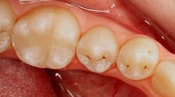
Identification of occlusal pit caries is aided by visual cues of white opacities at the entrance of grooves and enamel roughening within the groove. Identifying these lesions may be aided by tools that employ light-induced fluorescence. Red laser light-based (655 nm wavelength) devices (DIAGNOdent Pen, KaVo) provide a number to indicate the amount of fluorescence reflected from bacterial byproducts, whereas blue light-based (400-450 nm wavelength) devices (DIAGNOcam, KaVo; CamX Spectra, Air Techniques) produce a map on the tooth of luminous intensity in areas where there is bacterial activity. Blue light-based devices are reported to be more accurate than red laser light-based devices.6 Radiographic evidence of occlusal caries is not observed until later stages of demineralization.
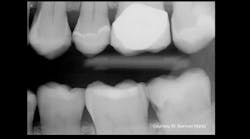
Due to contact with neighboring teeth, identification of interproximal caries is typically performed using radiographs. Identification of incipient enamel lesions can be aided with artificial intelligence (AI) tools (OverJet AI, Pearl), which have been shown to improve sensitivity for identification from 64% without AI to 75% with AI.7 Interproximal caries can also be identified with near-infrared transillumination (DIAGNOcam, KaVo; VistaCam, Durr Dental), which demonstrates caries as dark areas in which higher porosity carious enamel absorbs more light.
Treatment of incipient enamel lesions
Prior to enamel cavitation, incipient enamel lesions should be treated with nonoperative techniques with the goals of arresting the caries process, remineralizing the lesion, and preventing the cavitation of the outer wall of enamel. Cavitation is assessed visually for smooth surface and occlusal caries lesions. Cavitation is deduced from radiographic extent for interproximal lesions. Studies have indicated that only 11%–15% of radiographic lesions confined to enamel are cavitated.8,9
Treatment of incipient lesions may involve disrupting the caries process and remineralizing or infiltrating the demineralized lesion. The cornerstone of any caries management plan involves reducing the frequency of intake fermentable sugars and mechanically removing bacteria-containing plaque from teeth.
Additional approaches to reduce the effects of bacteria-mediated caries process is to reduce the bacteria present in the mouth. Various antibacterial products have been suggested, including xylitol, silver (Nano Silver Mouthrinse, Elementa), povidone iodine (Povi-One, Elevate Oral Care), and sodium hypochlorite (Treatment Rinse, CariFree). A relatively new approach is to provide probiotics, which contain strains of oral bacteria identified in patients with low caries incidence (Prodentis, BioGaia; Oral Health Probiotic, Bristle Health).
Remineralization of existing white spot lesions can be performed with fluoride toothpaste or varnish. Fluoride remineralization provides the benefit of replacing hydroxyapatite crystal with fluorapatite crystals, which are more resistant to future demineralization.10 Silver diamine fluoride is typically used for cavitated lesions; however, there is some evidence that it may also be effective for noncavitated lesions.11,12
Remineralization can also be performed with hydroxyapatite toothpaste or pastes with amorphous calcium phosphate in a casein phosphopeptide carrier (MI Paste, GC).13 A new concept in remineralization is a product (Curodont, vVardis) containing a peptide building block to direct calcium and phosphate in saliva to form hydroxyapatite crystals.14 Alternatively, a caries lesion may be infiltrated with resin (Icon, DMG) to arrest its progress.15
In summary, clinicians should be vigilant about detecting early signs of caries and apply nonoperative approaches to arrest these lesions in order to prevent surface cavitation and lesion progression.
Editor's note: This article appeared in the May 2025 print edition of Dental Economics magazine. Dentists in North America are eligible for a complimentary print subscription. Sign up here.
References
1. Takahashi N, Nyvad B. The role of bacteria in the caries process: ecological perspectives. J Dent Res. 2011;90(3):294-303. doi:10.1177/0022034510379602
2. Guo Z, Guillen DP, Grimm JR, Renteria C, Marsico C, Nikitin V, Arola D. High throughput automated characterization of enamel microstructure using synchrotron tomography and optical flow imaging. Acta Biomater. 2024;181:263-271. doi:10.1016/j.actbio.2024.04.033
3. DeRocher KA, Smeets PJM, Goodge BH, et al. Chemical gradients in human enamel crystallites. Nature. 2020;583(7814):66-71. doi:10.1038/s41586-020-2433-3
4. Frank RM. Structural events in the caries process in enamel, cementum, and dentin. J Dent Res. 1990;69 Spec No:559-66; discussion 634-6. doi:10.1177/00220345900690S112
5. Kidd EA, Fejerskov O. What constitutes dental caries? Histopathology of carious enamel and dentin related to the action of cariogenic biofilms. J Dent Res. 2004;83 Spec No C:C35-8. doi:10.1177/154405910408301s07
6. Macey R, Walsh T, Riley P, Glenny AM, Worthington HV, Fee PA, Clarkson JE, Ricketts D. Fluorescence devices for the detection of dental caries. Cochrane Database Syst Rev. 2020;12(12):CD013811. doi:10.1002/14651858.CD013811
7. Mertens S, Krois J, Cantu AG, Arsiwala LT, Schwendicke F. Artificial intelligence for caries detection: Randomized trial. J Dent. 2021;115:103849. doi:10.1016/j.jdent.2021.103849
8. Urzúa I, Cabello R, Marín P, et al. Detection of approximal caries lesions in adults: a cross-sectional study. Oper Dent. 2019;44(6):589-594. doi:10.2341/17-314-C
9. Pitts NB, Rimmer PA. An in vivo comparison of radiographic and directly assessed clinical caries status of posterior approximal surfaces in primary and permanent teeth. Caries Res. 1992;26(2):146-152. doi:10.1159/00026150008
10. Simmer JP, Hardy NC, Chinoy AF, Bartlett JD, Hu JC. How fluoride protects dental enamel from demineralization. J Int Soc Prev Community Dent. 2020;10(2):134-141. doi:10.4103/jispcd.JISPCD_406_19
11. Polacek J, Malhi N, Yang YJ, Scully AC, Soki FN, Boynton JR. Silver diamine fluoride and progression of incipient approximal caries in permanent teeth: a retrospective study. Pediatr Dent. 2021;43(6):475-480.
12. Braga MM, Mendes FM, De Benedetto MS, Imparato JC. Effect of silver diamine fluoride on incipient caries lesions in erupting permanent first molars: a pilot study. J Dent Child (Chic). 2009;76(1):28-33.
13. Raphael S, Blinkhorn A. Is there a place for tooth mousse in the prevention and treatment of early dental caries? A systematic review. BMC Oral Health. 2015;15(1):113.
14. Kind L, Stevanovic S, Wuttig S, et al. Biomimetic remineralization of carious lesions by self-assembling peptide. J Dent Res. 2017;96(7):790-797. doi:10.1177/0022034517698419
15. Chatzimarkou S, Koletsi D, Kavvadia K. The effect of resin infiltration on proximal caries lesions in primary and permanent teeth. A systematic review and meta-analysis of clinical trials. J Dent. 2018;77:8-17.doi:10.1016/j.jdent.2018.08.004
About the Author

Nathaniel Lawson, DMD, PhD
Dr. Lawson is an associate professor and the director of the division of biomaterials at the University of Alabama at Birmingham School of Dentistry. He graduated from UAB School of Dentistry in 2011 and obtained his PhD in biomedical engineering in 2012. He has lectured internationally, served as an investigator on numerous research grants, and published extensively on dental materials. He also works as a general dentist in the UAB Faculty Practice.
Updated September 25, 2023

Silvia Rojas-Rueda, DDS
Silvia Rojas-Rueda, DDS, is a pediatric dentist from Colombia currently pursuing a Master of Science and residency in dental biomaterials at the University of Alabama at Birmingham. With experience in clinical care and academic research, her work explores material performance, esthetics, and digital applications in modern restorative dentistry.