Failure to graft fresh extraction sockets can lead to costly second surgeries
OsteoGraf®/LD-300 reduces risk of ridge resorption in extraction sites.
A special feature presented with the cooperation of CeraMed Dental, LLC
Philip Gruell, DDS
Today`s dentistry is very different than that of years ago. Today`s patients demand not only quality dentistry but insist on cosmetically pleasing restorations. As dental practitioners, it is our responsibility to provide the best clinical work possible and deliver both conventional and/or implant-supported prosthetics that closely resemble natural dentition.
The challenge arises when patients exhibit less-than-normal oral anatomy, particularly of the residual ridge. Residual ridge resorption (RRR) has been well-documented in the literature dating back to 1935 with Deebach`s(1) article on the healing process following the removal of teeth. For more than 60 years, clinicians and scientists alike have reported on the consequences resulting from incomplete bone fill of the alveolar socket after tooth extraction. Pietrokovskia and Massler(2) (University of Illinois) studied the morphologic changes that take place after a tooth is extracted and the upward plus inward resorption pattern of the alveolar maxilla. This resorption process can make the commitment of a natural-looking restoration for our patients more difficult to achieve. Yet, many of us do not realize the necessity for grafting fresh extraction sockets for preservation of the alveolar process. Maintenance of the ridge form is of utmost importance if esthetics dictate the degree of satisfaction for the patient.
Tooth extraction is one of the most common surgical procedures performed in dental offices. For decades we have thought that socket healing included bone formation and bone fill. In 1960, Amler(3) reported the human alveolar socket healing sequela in undisturbed extractions. He explains that mineralization is apparent at day 18, but by 38 days, the laminadura had lost its definitive outline. At this stage the alveolar crest appears to undergo a slight resorption, which progressively increases. The bone fill that occurs in extraction sites falls short of the original crestal height and width and continues to degrade over time.
Misch et al. can be quoted as saying, "During the first year after tooth loss, 40 - 60% of the width of the alveolar ridge resorbs after tooth extraction(4)." The resorption and collapse of the hard and soft tissue architecture increases the complexity of the restorative treatment plan for the doctor and has a dramatic negative effect on the esthetics of the final prosthesis. To prevent this complication, grafting extraction sockets with alloplastic materials has become a routine part of extraction surgery in my office.
Studies provide the clinical rationale for grafting fresh extraction sockets as preventive care. Preserving the alveolar ridge offers patients choices in their restorative treatment plans, including endosseous implants(5). With a variety of alloplasts commercially available, the composition of the material should be evaluated for its suitability for the clinical indication. In an extraction socket, an inexpensive, resorbable calcium phosphate material can be used such as OsteoGraf®/LD-300 [CeraMed Dental, L.L.C., Lakewood, CO]. It is a pure form of calcium phosphate, highly porous and resorbable. It provides a readily available source of calcium that is necessary for mitogenesis and can be used successfully for ridge preservation.
The formation of new bone in a four- or five-wall defect, such as an extraction socket, will occur by direct attachment to the old bone spicules with preliminary resorption of the old bone. Bone taken from a site previously grafted with OsteoGraf/LD-300 was histologically confirmed by a core specimen taken at 16 months. Histology (Michael Rohrer, DDS, University of Oklahoma) showed relatively dense trabeculae with areas of bone marrow(6). Importantly, no evidence of graft material was seen, supporting the remodeling to bone. Resorbable calcium phosphates are biocompatible, they provide calcium for bone formation, and, since bone completely surrounds the defect, preserve the alveolar ridge form after tooth extraction. This preservation eliminates the need for a re-entry surgical procedure to repair and/or augment any potential deformity of the residual ridge.
OsteoGraf/LD-300 is supplied in sterile 1- and 3- gram vials. The unique vial interconnect accepts three different styles of syringes (straight, curved, or beveled tip). The unfilled syringe barrel is inserted into the vial connector, and the desired amount of OsteoGraf/LD-300 is transferred from the vial into the syringe. (Most extraction sockets require less than 1/2 gram of OsteoGraf/LD-300.) The nylon filter cap is replaced on the syringe and either sterile saline solution or sterile water can be drawn up through the filter cap to thoroughly wet the material. Once hydrated, depressing the syringe plunger expels the excess wetting agent. The filter cap is then removed and the hydrated OsteoGraf/LD-300 can be expressed into the prepared socket.
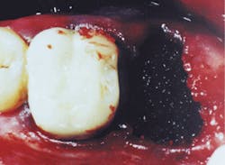
The graft material should completely fill the socket to the height of the alveolar crest (Figure 1). The placement of the graft material is accomplished in approximately 5 mm increments to ensure a uniform density throughout the graft. Upon placement, each of these layers is condensed into the socket firmly, but loose enough to maintain the blood supply throughout the graft. The close approximation of the grafting material to the fresh, clean bony wall optimizes the osteogenic potential of the site.
An added benefit of this material is the reautoclavable vial feature. As clinicians, we dispose of many products that are not intended for multipatient use. The OsteoGraf vials can be returned to a steam autoclave up to three times after the initial use, making this product even more economical for the clinician.
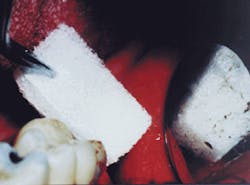
When grafting socket(s), primary closure cannot always be achieved. In these cases, epithelium begins to proliferate from the margins of the wound and migrate across at a rate of 0.5 mm per day. It can be assumed that in a period of 10 - 14 days enough granulation tissue will have been formed to cover the socket(s) and contain any graft material present. The period of concern is immediately following the placement of the graft until the time the natural healing response has covered the socket(s). Particulate graft material in itself will help prevent epithelial down-growth into the extraction socket, but a membrane is needed to aid in containing the graft material. This is in contrast to guided tissue regeneration (GTR), in which a true barrier membrane material is specifically designed to prevent epithelial down-growth for a longer period of time. Gelfoam® Sterile Sponge (Pharmacia & Upjohn, Kalamazoo, MI) is a medical device intended for application to bleeding surfaces as a hemostatic. Surface-acting hemostatic devices, such as Gelfoam, arrest bleeding by providing a mechanical matrix that facilitates clotting. Due to its bulk, Gelfoam slows the flow of blood, protects the forming clot, and offers a framework for deposition of the cellular elements of blood. It is a porous, pliable, sponge-like material that can be cut to size without fraying or losing its integrity. It is easily placed over fresh extraction sites, regardless of the size or configuration of the socket. Applied dry, Gelfoam can be compressed to adapt firmly to the socket walls (Figure 2). According to the manufacturer, Gelfoam will break down in approximately one week and is absorbed completely in four to six weeks. Gelfoam seems to maintain control of the site long enough for epithelization to contain the graft. It offers a protective cover and provides structural support for the reparative process. It is an inexpensive way to achieve containment of the OsteoGraf/LD-300 in the socket walls.
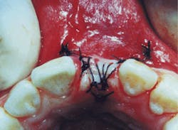
A simple suture is secured with minimal tension over the grafted socket to avoid loss of the OsteoGraf/LD-300 and the Gelfoam (Figure 3). Standard postoperative instructions are given to the patient, and an appointment is scheduled for suture removal in 10 days.
Since it is unlikely to be covered by insurance, socket grafting must be presented to patients as a service and a preventive measure that will result in a better restorative procedure and/or a more pleasing cosmetic result. Few patients refuse grafting of fresh extraction sockets when they realize the benefits and possible alternative to more complex restorative procedures. By implementing this service in my practice, I have improved the quality of the relationships with my patients and enhanced my practice productivity. This fee-for-service procedure is inexpensive to the practice. In most cases, less than 1/2 gram of OsteoGraf/LD-300 will graft a single socket, which equates to less than $20 in material costs for the practice. It requires minimal additional chair time, and my fees average $80-$100 for the graft. Grafting fresh extraction sockets is extremely valuable to the patient and has become a standard of care in my practice.
For several years, I have practiced this method of ridge preservation. This practice is more important now than ever before. With the enormous concern for esthetics and interest in cosmetic dentistry, clinicians are denying their patients the optimal treatment if they are not grafting every extraction site.
Many of my patients are referred to my practice for cosmetic consultations. They are unhappy with their existing prosthesis and want a more natural-looking, functional restoration. It has become obvious to me that if the socket(s) had been grafted at the time of the extraction, fewer of these patients would require the additional grafting and/or additional surgeries they are now facing to restore the contour of the alveolar ridge. The principles mentioned above are exemplified by the two cases discussed in the related articles.
Although predictable and dependable, the surgical procedures shown in the two cases on pages 96-97 shouldn`t be necessary. Grafting sockets at the time of extraction reduces the risk of residual ridge resorption, and often eliminates the need for more extensive grafting in the future. By utilizing the OsteoGraf/LD-300 as a bone filler in fresh extraction sockets, the risk of ridge resorption is minimized. Without grafting, bone will continue to resorb during the life of the patient, making it difficult to maintain fit, function, and esthetics of any prosthesis. The time involved to graft fresh extraction sites is minimal compared to the time required to regraft and regenerate new bone to an atrophied ridge.
About the author
Dr. Philip Gruell graduated from the University of California and received his dental degree from the University of the Pacific, San Francisco, California. He held faculty positions in the departments of Operative and Fixed Prosthetics before entering full-time private practice in Alameda, California. He is a Diplomate in the International Congress of Oral Implantology, a member of the Academy of Osseointegration, the Academy of General Dentistry, and the Academy of Cosmetic Dentistry. Dr. Gruell is the co-director of two San Francisco-area implant study groups, and maintains a private practice emphasizing implant and cosmetic dentistry. Dr. Gruell can be contacted at his Alameda, Calif., office at (510) 523-7813.
This article was presented with the cooperation of CeraMed Dental, LLC. Additional information on OsteoGraf/LD-300 can be obtained by contacting a technical representative at CeraMed Dental at (800) 426-7836.
References
1. Deebach, R.F.: Healing Process Following the Removal of Teeth Where Injury Greater Than the Sustained in Simple Extraction Is Involved, Northwestern Univ. D. Res. & Grad. Quart. Bull. 35:4, 1935
2. Pietrokovski, J., Massler, M.: Alveolar Ridge Resorption Following Tooth Extraction, Journal of Pros. Dent, Vol. 17, No. 1, January 1967.
3. Amler, M., Johnson,P., Salman,I.: Histological and Histochemical Investigation of Human Alveolar Socket Healing in Undisturbed Extraction Wounds, Journal of ADA, Vol. 61, July 1960.
4. Misch, C. Dietsh,F.: Bone Grafting Implants in Implant Dentistry, Implant Dentistry, Vol. 2, No. 3, 1993
5. Klute-Murray, V.: Anterior Ridge Preservation and Augmentation Using Synthetic Osseous Replacement Graft, Compendium, 19(1):69-77, 1998
6. Murphy, F.: OsteoGraf/LD-300 and Extraction Sites, CeraMedia, 1st Quarter - 1997
Photography courtesy of Christine Lyddon-Murray, RDA
Figure 1
Figure 2
Figure 3
Case #1
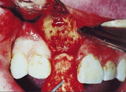
A 17-year-old male was referred to me 11 months following traumatic avulsion of tooth #8. At that time, he was wearing a stayplate as a cosmetic replacement for the missing anterior tooth. Although told by his former dentist that he was ready for an implant-supported crown, it was apparent from clinical examination that significant resorption of the alveolar ridge had already taken place (Figure 4). Therefore, ridge augmentation was necessary to restore the facial contour to its original dimension before an implant could be placed.
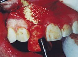
Mid-crestal and vertical releasing incisions were made to provide access to the remaining facial bone. A #6 round bur was used to fenestrate the recipient site to provide vascularity for the graft (Figure 5). Prior to placement of the grafting material, a porous resorbable membrane, BioMend® (SulzerMedica, Carlsbad, CA), was contoured and secured in position under the edge of the palatal tissue with a 4-0 Vicryl® (Ethicon, Summerville, NJ). A composite mixture of bovine-derived bone mineral, OsteoGraf® N-300 and DFDBA (AlloSource, Denver, CO) was hydrated with a dilute solution of tetracycline and sterile water. The graft mixture was uniformly packed into the prepared recipient site (Figure 6). The opposite free end of the membrane was positioned under the facial tissue flap to contain and shape the particulate graft. The previously released facial tissue was then reapproximated, and a close interlocking closure was made with the Vicryl suture (Figure 7). In four months sufficient bone will have been regenerated to allow for placement of an endosseous implant.
Figure 4
Figure 5
Figure 6
Figure 7
Case #2
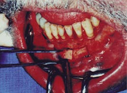
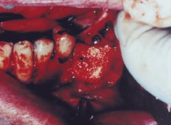
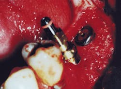
A more aggressive surgical approach was necessary to regain the height and width lost after extraction of tooth #18 on this 57-year-old male. The patient was unwilling to wear a removable partial denture, but was concerned about maintaining his oral health and function. The severity of this defect was best addressed by grafting with a combination of autogenous cortical bone and particulate bovine bone mineral. Two cortical segments were removed from the symphysis region (Figure 8). They were shaped and contoured for careful adaptation to the recipient site. The area of the defect was trephinated with a #6 round bur, and the donor bone was rigidly fixated using 1.5 mm titanium screws. Autogenous marrow that had been harvested from the chin was mixed with OsteoGraf N-300, and used as a mortise around the cortical blocks (Figure 9). The donor site was returned to full contour with OsteoGraf N-300. To reduce the risk of soft tissue invasion, both areas were covered with BioMend resorbable membranes prior to routine closure with Vicryl sutures. After six months, sufficient bone had been regenerated to accept two 4.3 mm Replace® implants (NobelBioCare, Yorba Linda, CA) (Figure 10).
Figure 8
Figure 9
Figure 10


