Implant esthetics
Cutting corners 'doesn't cut it'!
W. Johnston Rowe, Jr. DDS, AAACD
Patient expectations of implant esthetics have increased dramatically in the last decade. Advances in 3-D imaging and tissue grafting allow for better planning and more precise implant placement than ever before. Technology combined with proper handling of today's restorative materials in the hands of a competent practitioner can allow esthetic implant results that are often mistaken for natural teeth.
Case presentation patient history and chief concern
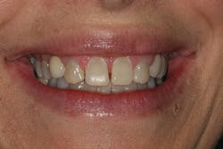
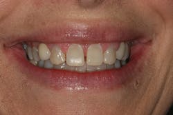
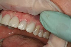
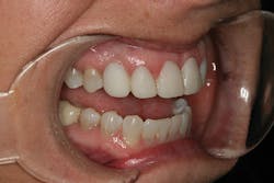
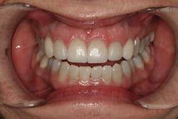
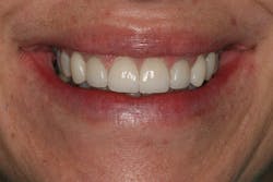
A 32-year-old white female presented for a cosmetic consultation. The patient reported that during her teenage years she had received orthodontic treatment to move her permanent maxillary canines mesially into the spaces that should have been occupied by her congenitally missing lateral incisors. She stated that her dentist, orthodontist, and oral surgeon had decided to place endosseous implants in the canine spaces due to the greater availability of bone in comparison to the bone available in the lateral positions (figure 1). As she grew older, she had become increasingly dissatisfied with the appearance of her smile and was seeking a more updated and esthetic solution (figure 2).
Figure 1
Figure 2
Treatment plan and phasing
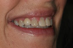
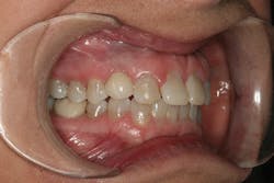
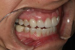
A comprehensive cosmetic, radiographic, and functional evaluation was performed.1 The patient was able to identify the major concerns leading to her dissatisfaction with her smile, including the presence of multiple diastemata, tooth proportion improprieties, unnatural emergence profile of her implant restorations, and her desire for a whiter smile (figure 3).2
Discussion occurred regarding the positioning of implant Nos. 6 and 11. The patient was made aware that the positioning of the implants would prove to be a guiding factor in the execution of her treatment plan. Additional surgical options were entertained and the patient refused these options. The patient chose to keep the implants in their current position and a treatment plan was established. The patient chose to pursue tray bleaching and replacement of the implant restorations with new custom abutments and porcelain crowns. A provisionalization stage was employed in order to retrain the soft tissue to develop a more natural emergence profile.3,4 Size, spacing, and color issues were to be addressed with the restoration of teeth Nos. 7 through 10 with porcelain laminate veneers.
Treatment
Diagnostic casts were used to fabricate a set of custom whitening trays. A two-week tray-whitening regimen (Opalescence 20% PF, Ultradent) was employed to enable the patient to whiten all of her natural teeth and begin to envision the shade she would wish to obtain in her final restorations.
A mounted diagnostic wax-up was fabricated to serve as a template for the planned restorations.5 Special attention was given to the contouring of the proposed restorations of the implant crowns. Having had the canines Nos. 6 and 11 orthodontically moved into the lateral positions (Nos. 7 and 10) also repositioned the osseous prominences of the canine eminentia mesially. Careful handling of the wax-up was necessary to plan for appropriate facial soft tissue support in the areas of Nos. 6 and 11 without providing an unnatural emergence or bulk of the restorations. A provisional stent and a reduction guide (SilTech, Ivoclar Vivadent) were then fabricated from the diagnostic wax-up.
The patient did not know the manufacturer or size of her implants, and efforts to locate her dental records from the surgeon who placed the implants were unsuccessful. Radiographic evaluation was employed to determine the type of implants placed prior to initiation of treatment. Use of reference information found on the website www.whatimplantisthat.com proved extremely helpful, as did the ability to use digital calipers on a study model to calibrate measurements of the implants in digital radiographs. The implants were determined to be Nobel Biocare Branemark NP 3.5 mm external hex endosseous implants (Nobel Biocare). Given this information, restorative components were ordered prior to initiation of treatment. This eliminated the potential for having to employ a trial-and-error process of ordering and trying in multiple types of fixtures once the existing abutments were removed.
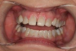
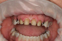
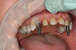
After approval of the diagnostic wax-up, profound anesthesia in the area of Nos. 6 through 11 was achieved (Septocaine, Septodont). Following sounding for bone to confirm adequate biologic width, laser soft-tissue recontouring in the areas of Nos. 6 through 11 was performed using an Er;Cr:YSGG soft- and hard-tissue laser (Waterlase, Biolase). Teeth Nos. 7, 8, 9, and 10 were prepared for conventional porcelain laminate veneers. Implant crowns were sectioned and removed from Nos. 6 and 11 (figure 4). To verify the radiographic implant identification, the abutments were removed and impression copings were tried in and radiographic seat was confirmed. The abutments were then replaced to serve as provisional restorative components. Screw access holes were blocked out and the provisional stent was used to fabricate provisional restorations on Nos. 6 through 11 (Luxatemp Ultra BL, DMG America) (figures 5, 6).
Figure 3
Figure 4
Figure 5
Figure 6
Figure 7
Figure 8
Figure 9
Figure 10
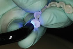
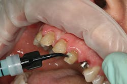
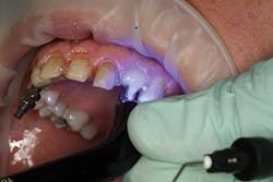
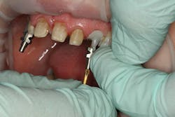
The provisionals were then removed and the gingival contact areas of Nos. 6 and 11 were modified with a flowable composite material to support the soft tissue and develop a more natural emergence profile3 (figures 7, 8). When appropriate support was achieved, the gingival contact areas of the provisional were polished to a smooth shine, free of rough areas. The provisionals were then trimmed, contoured, stained with tints, polished, and glazed (Luxaglaze, DMG America) prior to seating with a clear resin cement (TempBond Clear, Kerr). The patient was given home-care information and instructed to return in two weeks to evaluate the provisional restorations and tissue response around the implant sites.
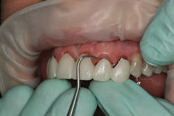
During the two-week evaluation appointment, the tissues were determined to be responding appropriately (figure 9). Had they not, the provisionals would have been removed and modified before reseating and appointing for another two-week evaluation. As the tissue in this case was responding as expected, the patient was appointed to return in another two weeks for master impressions.
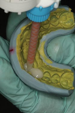
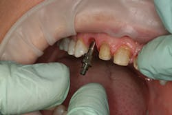
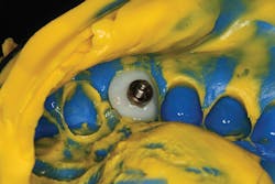
At the beginning of the impression appointment, the provisionals were approved for fit and esthetics with a particular focus on emergence profile of the implant provisionals. Profound anesthesia was achieved and the bisacrylic provisionals were removed. The preparations were cleaned with chlorhexidine (Consepsis, Ultradent) and the abutments were once again removed (figure 10). The open-tray impression copings were then tried in, their seat confirmed radiographically, and then modified into custom impression copings with flowable composite (figures 11-14). Once appropriate tissue support was confirmed, the open-tray master impression was made with a heavy and light body wash technique using vinyl polysiloxane impression material (Honigum, DMG America) (figure 15). Following the master records, the provisional abutments and bisacrylic provisionals were replaced until the seat appointment.
Figure 11
Figure 12
Figure 13
Figure 14
Figure 15
Figure 16
Figure 17
Figure 18
Figure 19
Careful technique and accurate records provided the lab with the information required to provide a result that replicated the contours of the provisional restorations. The seat appointment proceeded smoothly with no adjustments (figures 16, 17).
Conclusion
This case clearly illustrates the benefit of proper tissue management and tooth contour. Modification of tissue support and tissue training were the key elements in developing natural implant results that took this patient from being dissatisfied to loving her smile. The additional time invested in planning and manipulating soft tissues was critical to the long-term success of this implant case (figures 18, 19).
References
1. Dawson PE. Evaluation, Diagnosis, and Treatment of Occlusal Problems. St. Louis, MO: Mosby; 1989.
2. Finlay SW. Contemporary Concepts in Smile Design: Diagnosis and Treatment Evaluation in Comprehensive Cosmetic Dentistry: A Guide to Accreditation Criteria. Madison, WI: American Academy of Cosmetic Dentistry; 2014.
3. Fradeani M. Esthetic Analysis: A Systematic Approach to Prosthetic Treatment: Volume 1. Chicago, IL: Quintessence Books; 2004.
4. Rufenacht CR. Fundamentals of Esthetics. Chicago, IL: Quintessence Books; 1992.
5. Magne P. Bonded Porcelain Restorations in the Anterior Dentition: A Biomimetic Approach. Chicago, IL: Quintessence Books; 2002.
W. Johnston Rowe Jr., DDS, AAACD, maintains a private practice dedicated to excellence in general, cosmetic, and complex restorative dentistry located in Jonesboro, Arkansas. He is an accredited member of the American Academy of Cosmetic Dentistry and currently serves as the chairman of the American Board of Cosmetic Dentistry. He also serves as an Accreditation Examiner for the American Academy of Cosmetic Dentistry. Dr. Rowe has been awarded fellowships in the International College of Dentists and the Pierre Fauchard Academy. He is a graduate of the University of Tennessee College of Dentistry and is a formally trained artist having graduated from Washington and Lee University with a Bachelor of Arts degree in studio art. Dr. Rowe enjoys sharing his passion for cosmetic dentistry materials and techniques, lecturing nationally and internationally, and can be contacted through his office at [email protected] or (870) 932-4126.
