Laser-assisted pontic preparation
Michael DiTolla, DDS, FAGD
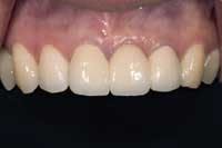
Despite the increasing availability of single-tooth implants, many patients choose to close edentulous spaces with fixed prosthodontics. In addition to other critical esthetic factors, pontic design can often make or break the final esthetic result. The use of an ovate pontic receptor site is of great value when trying to create a natural maxillary anterior fixed bridge. This article will illustrate the creation of an ovate pontic receptor site with the use of an ErCr:YSGG hard and soft tissue laser. I have found this to be the easiest, most atraumatic way to create these types of pontic receptor sites.
The ovate pontic receptor site is a depression or socket created in the soft tissue that allows the cervical aspect of the pontic to emerge from it, making it appear to emerge from the alveolar ridge. Modified ridge lap pontics have the disadvantage of usually appearing too long when compared to adjacent or contralateral teeth. There are two scenarios that can occur prior to creating an ovate pontic receptor site: the patient with the tooth that still needs to be extracted, and the patient with already established edentulous space.
In the case of the tooth that needs to be extracted, treatment is relatively straightforward, predictable, and is typically an esthetic success. The big advantage in a situation like this is the presence of the interdental papilla prior to extraction that will allow the clinician to achieve a superior esthetic result. The most predictable way to treat this type of case is with a lab fabricated provisional restoration. The laboratory "extracts" the tooth to be removed on the model, and then "sockets" the model to mimic what the extraction site will look like. It is preferable to have the tissue side of the pontic longer rather than shorter since there is no chance of biologic width violation in this case..The lab should attempt to have 4-5 mm of pontic extend into the extraction site apical to the free gingival margin. The key is to preserve the papilla during the extraction procedure and to fill the extraction site with the provisional pontic as soon as possible. The time-honored practice of having the patient bite down on a 2x2 gauze to stop the bleeding is contraindicated in this procedure. Since the tooth has been extracted, the interdental papillae have no support until the provisional is cemented, and by biting on the gauze, the patient will most likely collapse the papilla. In order to preserve as much of the facial and interproximal bone as possible, it behooves the clinician to strive for an atraumatic extraction, with the use of more conservative instruments, such as periotomes.
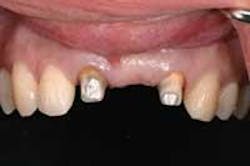
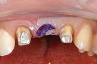
In the case of a healed edentulous site, use the lab fabricated provisional to help guide you with your pontic site development. The tissue side of the pontic is marked with a color transfer applicator, and the provisional is set until it contacts tissue. Remove the bridge, and with the Waterlase on the soft tissue setting, begin to develop the pontic site by removing the tissue where the ink is present. When the ink has been removed, reseat the provisional and continue to sculpt the tissue wherever ink is present. When you reach the point that all 3 mm of the tissue side of the pontic are apical to the free gingival margin, and the bridge seats without blanching the socket site, you are finished with the soft tissue sculpting. However, biologic width remains a concern. Insert a periodontal probe into the deepest part of the pontic site and push it into the tissue until it contacts bone. If there is 2 mm or more tissue remaining on the crestal bone, you are ready to cement the provisional bridge. If there is less than 2 mm of tissue remaining, it is necessary to remove enough crestal bone to allow for the 2 mm of gingiva between the bone and the pontic. With the Waterlase at the hard tissue setting, 1 mm of bone was conservatively removed. Ensure that you leave a minimum of 2 mm of space between the tissue side of the pontic and the crestal bone to allow the soft tissue to fill this space.
If you are confident about your success with this procedure, you can take final impressions at this appointment as well. Otherwise, the patient is appointed post-operatively in seven days, at which time the provisional bridge is removed, the pontic site is evaluated and final impressions are taken.
Dr. Michael DiTolla is director of clinical research and education at Glidewell Labs in Newport Beach, Calif., where he also teaches over-the-shoulder courses on topics such as esthetic restorative dentistry. Dr. DiTolla also teaches a two-day, live-patient, hands-on laser-training course that emphasizes diode and erbium lasers. In addition, he teaches a two-day, hands-on digital photography course emphasizing intraoral and portrait photography, and image manipulation. More information on these and other courses can be found by email at [email protected] or by calling (888) 535-1289.


