Incorporating CBCT into your dental practice
Ara Nazarian, DDS, DICOI
Integrating technology such as cone beam computed tomography (CBCT) within your practice enhances your ability to accurately diagnose, properly present, and effectively deliver dental treatment. With today’s CBCT systems, we are seeing increased speed, clarity, and quality of images, reduced levels of radiation exposure, and deeper integration with treatment planning software.
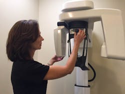
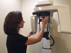
Now that I have this technology in my office (CS 8100 3D, Carestream Dental; figure 1), I totally understand these advantages and much more. However, before I purchased and implemented this technology, I wanted to carefully contemplate the pros and cons and my return on investment with this type of equipment. After examining these considerations, I determined that having a CBCT imaging unit readily available in my practice could help me eliminate loss of revenue, increase case acceptance, and improve patient care.
Figure 1: The CS 8100 3D
How many times have we heard from our patients, “Doc, can’t you just do this root canal?” or “take out this tooth”? Patients feel comfortable with their primary dentists and don’t want to be referred out unless they absolutely need to be. But without the technology that allows you to precisely visualize the vertical fracture or missed canal that is causing the patient discomfort, you have little choice. However, with the right technology, you are better positioned to diagnose and address the situation within your comfort level, thus satisfying the patient and keeping that stream of revenue inside your practice. And even in those situations where you are not comfortable with executing a particular procedure, an accurate diagnosis and precise instruction or recommendation to the correct specialist still builds and enhances your patient’s confidence in your abilities.
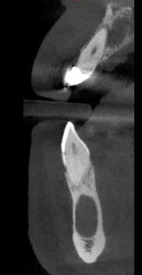
Another example is the new emergency patient who presents to your practice with severe pain. He or she had been telling the previous dental provider several days or weeks earlier that a particular tooth was causing discomfort; however, the dental provider saw no indications of tooth decay, infection, fracture, missed endodontically treated canals, or pathosis (figure 2) since he or she did not have a CBCT system. But, since you have the right tool for the job, you can zero in on the cause of pain and address it immediately.
Figure 2: Upper periapical lesion, lower possible odontogenic keratocyst
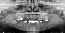
When a new patient presents to my practice for an initial oral examination, I usually obtain a 2-D or 3-D image, depending on what he or she requires in terms of treatment. In other words, if a routine new patient presents to the practice, then we obtain a 2-D panoramic image. Due to its low dose and simplicity, digital 2-D panoramic imaging remains an indispensable tool for the majority of dental practices. And whether you are a general practitioner or a specialist, a compact multifunction system with multiple fields of view can cover all your routine panoramic needs (figure 3).
Figure 3: An example of a panoramic image produced by a CBCT system
On the other hand, 3-D imaging can enhance the standard of care for endodontics, implants, oral surgery, and daily procedures of the general practice. Selectable fields of view give you what you need for a faster, more accurate, task-specific diagnosis. You can obtain your desired image while controlling image size, resolution, region of interest, and dose. In terms of dose, the safety of the patient is always the highest priority.
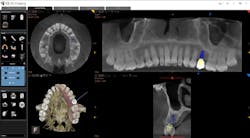
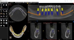
Whether it is an implant consultation for a single implant (figure 4), multiple implants, or full-mouth reconstruction, advanced 3-D software, such as the CS 3D Imaging software, offers you the ability to virtually plan the implant placement in regard to bone height, width, and density with respect to anatomical structures. Once planned, this information is reviewed with the patient in a clear and concise way that is easily understood (figure 5). In my experience, patients often state that they didn’t know that this type of technology was even possible within a dental office. Because of these reasons and more, I have found that case acceptance has gone up exponentially.
Figure 4: Planning an implant in CBCT software
There is no doubt in my mind that the incorporation of CBCT into your practice improves your patient care because it allows you to see a vast array of anatomical structures and conditions that may have been previously overlooked. With only a 2-D image of an area—whether it is for an endodontic procedure, removal of wisdom teeth, or placement of an implant—you are somewhat limited. However, with CBCT you can accurately identify an accessary canal in a tooth before to a root canal, a rotation of an impacted tooth prior to extraction, or pathology in a particular segment of bone.
Figure 5: Virtual planning of immediate implant placement
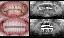
In today’s practice, dental professionals should have imaging that can satisfy all their diagnostic needs. CBCT is an investment that generates a strong return, especially when it covers nearly every routine situation as well as more advanced clinical situations (figure 6). With the systems currently available, you can cost-effectively take the essential first step with 2-D panoramic views and investigate in depth with powerful 3-D imaging. As a result, you can offer and perform more procedures in your practice, improving the overall level of patient care.
Figure 6: A full-mouth implant reconstruction achieved with CBCT
