Determining gingival margin position

Refine your esthetic workflow by mastering smile design from incisal edge to gingival margin. This step-by-step guide shows how to use clinical photos, ideal tooth proportions, and diagnostic tools to create natural, harmonious smiles.

Key Highlights

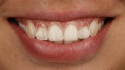
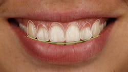
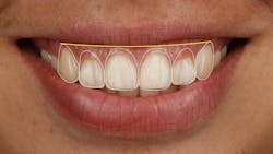
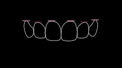
  • Determine ideal tooth proportions: Use the established incisal edge position to design tooth outlines with an 80% height-to-width ratio, ensuring the incisal curve follows the lower lip for a natural esthetic flow.

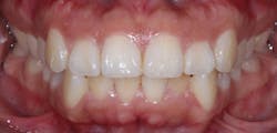
  • Align gingival margins for balance: Aim for centrals and canines on the same level, with laterals slightly coronal, maintaining symmetry and natural gingival display.

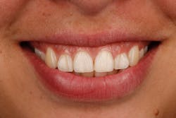
  • Evaluate gingival display and diagnosis: Identify “gummy smiles” (4 mm or more of gingival show) and use diagnostic systems like Global Diagnosis to plan necessary surgical or orthodontic adjustments.

In our previous article, we talked about the importance of the repose photo and determining incisal edge position. The next question is: Where do we go from here?

Once we’ve determined the incisal edge position from the repose photo, we can move to a smile photo or retracted anterior photo (figures 1 and 1a). Let’s walk through the step-by-step.

Using the proposed incisal edge position, we can draw in an ideal tooth outline on our smile or retracted anterior photo (figures 2 and 2a). There are many tools to help with this design; we often use Trios Smile Design or simply Keynote or PowerPoint. We like to use an 80% height-to-width ratio utilizing the existing width of the tooth, though studies show anywhere from 70% to 90% is considered esthetic.1,2 If we have space between the anterior teeth we are trying to fill, we use a 10 mm length and 8 mm width as a starting point. Draw in the laterals and canines as well, even the premolars if you’d prefer (figure 2b). This is especially helpful in the full smile photo. We’d like to see the incisal edges of our anterior teeth follow the curvature of the lower lip (figure 3).

Ideally, in a full smile, we’d like to see the gingival margins of the maxillary anterior teeth exist at the margin of the upper lip (figure 4). Natural smiles tend to show some gingival tissue as opposed to artificial smiles where you only see teeth. Think about this when you design your dentures and hybrids and create a more natural look. The most ideal margin positions show the canines and centrals to be on the same level with the lateral incisors slightly coronal to this line (figure 5). In the real world, we can’t always achieve this, so try to keep the gingival levels of the centrals equal and as much symmetry between laterals and canines as possible.

When we draw these ideal tooth shapes, we may find that the new gingival margins will require gingival surgery or tooth movement to achieve. One question we can ask is, “Does this patient have a gummy smile?” We’d consider 4 mm or more of gingival display as “gummy.” If the patient does show more than 4 mm, we need to consider the diagnosis. This will help us determine how we get to the new gingival margin. A system like Global Diagnosis by Bill Robbins and Jeff Rouse is a great way to arrive at this diagnosis. More on that in future articles.

We hope this provides a great starting point for looking at your cases! 

Editor's note: This article appeared in the November/December 2025 print edition of Dental Economics magazine. Dentists in North America are eligible for a complimentary print subscription. Sign up here.

References

  1. Chu SJ, Tan JHP, Stappert CFJ, Tarnow DP. Gingival zenith positions and levels of the maxillary anterior dentition. J Esthet Restor Dent. 2009;21(2):113-120. doi:10.1111/j.1708-8240.2009.00242.x
  2. Magne P, Gallucci GO, Belser UC. Anatomic crown width/length ratios of unworn and worn maxillary teeth in white subjects. J Prosthet Dent. 2003;89(5):453-461. doi:10.1016/S0022-3913(03)00125-2

About the Author

Christopher D. Mazzola, DDS

Christopher D. Mazzola, DDS

Christopher D. Mazzola, DDS, graduated from the University of Michigan Dental School in 2009. He is a member of the visiting faculty of the Pankey Institute in Key Biscayne, Florida, where he teaches “Essentials 3: Restorative Integration of Form and Function” and “Mastering Dental Photography.” He also recently joined The Dental Advisor team as an evaluator. He practices restorative dentistry in Traverse City, Michigan, in a large fourth-generation practice. He is a member of the Academy of General Dentistry and the American Academy of Cosmetic Dentistry. Contact him on Instagram @christopherdmazzoladds and visit his website at traversedental.com.

Charlie Ward, DDS

Charlie Ward, DDS

Charlie Ward, DDS, graduated from University of Maryland Dental School in 2008. He is a member of the visiting faculty of The Pankey Institute in Key Biscayne, Florida, where he teaches the Mastering Aesthetics Restorative Dentistry and Dental Photography courses. He also recently joined the faculty for Align Technologies. He practices restorative dentistry in Baltimore, Maryland, with his partner Dr. Devon Conklin, and his wife and periodontist Dr. Melody Ward. He is a member of the Academy of General Dentistry and the American Academy of Cosmetic Dentistry.

Sign up for our eNewsletters
Get the latest news and updates