Tooth lightening A new concept for maximizing surface esthetics
By Professor Laurence J. Walsh
With the current strong levels of interest by patients in reducing the aging–related yellow shades within their teeth by bleaching, it is surprising that greater attention has not been directed to other means of reducing yellowness. This paper looks at the concept of "tooth lightening" using simple in–office and supporting at–home measures, as opposed to "tooth whitening" using peroxides.
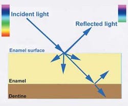
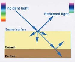
The concept is based on exploiting a number of well–established optical properties of teeth, enamel, and water under visible light conditions. These are presented step–wise in the accompanying tables and figures. The scientific foundations of the tooth lightening concept rest largely on altering the short wavelength (blue) scatter of enamel and reducing its transmission of yellow light, although there are minor accompanying changes such as reduced red absorption which also occur.
The clinical stages in tooth lightening are as follows. First, the reflection of light and the backscatter of the shorter wavelengths of light are enhanced by a gentle microabrasion procedure (ADA Item 116) using 37% phosphoric acid etching for 20 seconds followed by gentle application of flour of pumice or graded abrasive pastes at low rotational speeds.
The etching step enhances subsequent subsurface mineral changes. Patients then apply GC Tooth Mousse Plus™ each night immediately before bed for at least two weeks.
The reduced yellow transmission and increased backscatter of blue light from the enamel will cause a subtle reduction in yellow.
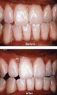
The procedure can precede in–office or at–home whitening treatments to establish optimal enamel properties and esthetics, or can follow other cosmetic treatments. A typical case is shown above. A variety of surface polishing agents can be used to maximize reflectivity without losing the natural surface contours.
Editor's Note: This article was reprinted with permission from the March/April 2008 edition of Australasian Dental Practice. References are available upon request.
Professor Laurence J. Walsh is the technology editor of Australasian Dental Practice magazine. He is also a noted commentator on and user of new technologies. Walsh is the head of the University of Queensland School of Dentistry.
OPTICS OF HUMAN TEETH
- The light that penetrates the surface of a tooth and enters it is refracted due to the fact that light travels faster in air than in water or in solid apatite materials.
- Tooth color is determined by the paths of light inside the tooth, and attenuation of light along those paths.
- Colors of natural teeth have a wide range (greater color space) than represented on existing shade guides, however yellow is the base color which causes the greatest distress when patients are asked to rate their own tooth color. The human eye is more sensitive to green and yellow light than to blue or red — because of the numerical difference in cones (color photoreceptors).
- Both reddish and yellowish colors of natural teeth tend to increase from the incisal to cervical, whereas translucency decreases. The middle and incisal thirds are the most visible during speech and in normal function, and thus yellowness here attracts the greatest attention.
- While a significant yellow fluorescence emission is excited from enamel under visible blue or ultraviolet lighting conditions, this makes little contribution to the perceived color of natural teeth under normal "white light" lighting conditions.
- Attenuation of light within the tooth is caused by several different factors, but primarily by scattering and absorption. Scattering of light disperses it, without changing the wavelength. Scattering can be forward or backwards in direction.
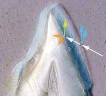
- Light is transmitted through enamel and dentine to the pulpal surface, with the light following the path of the enamel prisms and dentinal tubules. Enamel and dentine together collect and distribute light within the tooth.
- Light bends when it moves through dentine, in other words, dentine is optically anisotropic. Moreover, light propagation in human dentine exhibits a strong directional dependence.
- It was originally thought that both enamel prisms and dentinal tubules acted as optical fibers, however it is now well established that the tubular structure is the predominant cause of scattering in dentine.
- Multiple scattering events occur within the dentine tubules due to its cylindrical structure (and to a lesser extent by peri–tubular collagen fibers), and not by fiberoptic effects, as had been previously assumed.
THE INFLUENCE OF WATER PRESENT IN THE ENAMEL
- Enamel contains 7% to 10% water by volume as one of its normal constituents, and this water affects its ability to both absorb and scatter light.
- For early carious lesions and other enamel conditions with increased subsurface water, longer wavelengths of yellow and red are preferentially reflected to the surface. This radiance change is mainly determined by the lack of mineral and the increased water content in these lesions.
- Even without the development of white spot lesions or altered enamel formation, microscopic subsurface water voids are common as enamel is never perfectly mineralized. These water–rich areas can undergo subsurface remineralization by repeated topical application of CPP–ACP (Recaldent) products.
- Restoring mineral to these water–rich areas should, therefore, change the scatter of short wavelengths of light and alter the appearance and color of the enamel surface.
- Micropolishing the enamel surface makes tooth shade lighter and less yellow, and this effect is enhanced upon air drying, which removes subsurface moisture.
- This provides a rationale for micropolishing in combination with subsurface remineralization.


