Reducing continuing problems with zirconia crowns
Key Highlights
- Occlusion: Minimizing laboratory spacing and allowing minor clinical adjustments can improve zirconia crown occlusion and reduce the time needed for opposing teeth to interdigitate.
- Cementation and contacts: Conventional glass ionomer cement can help reduce marginal leakage and debonding, while requesting approximately 25 microns of additional contact contour can address loose contacts.
- Color and esthetics: Internal staining of zirconia in the green state can provide more stable color while eliminating the need for a feldspathic ceramic surface layer.
Each month, Dr. Gordon Christensen answers a question from readers about everyday dentistry.
Q: Overall, I am liking zirconia crowns, but in my opinion, they have some limitations. Some of the challenges I am seeing from various labs—not just from one lab—are:
- Usually zirconia crowns don’t interdigitate with the opposing teeth when they come from a lab. In other words, they are visibly not in occlusion.
- Often the contact areas are loose.
- I have had some crowns come off after short-term service in the mouth, and I see visible leakage and staining on the inside of the crown.
- Sometimes the contact areas are rough, and some catch floss.
- Usually the color is off by a shade or two.
- And there are a few other less frequently occurring issues that bother me.
Are there ways to reduce or eliminate these problems?
A: Yes, there are ways to reduce or eliminate the challenges you stated. I will discuss them one by one and include a few you may not have noticed.
Our organization, Clinicians Report Foundation (CR), has the largest long-term in vivo study of which I am aware. The answers I will provide for you have been seen in CR research and observed by many of our mentors (teachers) and evaluators.
The following suggestions will decrease or eliminate the challenges you mentioned.
Related: Solving zirconia occlusion challenges
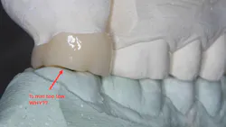
Crowns too low and overall anatomy from a crown library (figure 1)
Most laboratories purposely leave the zirconia crowns out of occlusion. Why? You know how difficult it is to smooth and polish zirconia. The labs do not want to inconvenience their clients by requiring them to adjust occlusion. Instead, the labs leave the crown too low, as much as 0.5 mm from occlusal contact with the opposing tooth. The clinical result caused by this action is that the newly crowned tooth and its opposing tooth (teeth) must extrude into occlusion. When they extrude, only a cusp or two will initially come into occlusion. At least a few months to several months are required for the opposing teeth and the new crown to interdigitate. At least a partial solution requiring less time for the extrusion is described below.
The solution: Advise your lab to space the crown minimally. I prefer about 25 microns of spacing (approximately one-half of the diameter of a human hair). Occasionally, you may have to slightly reduce a spot or two on the crown cusps or opposing tooth (teeth) to produce more optimum occlusion.
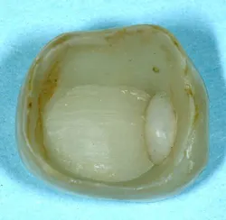
Staining on the intaglio surfaces of the crown or caries on margins (figure 2)
The margins of milled zirconia crowns have been shown by research to be open, up to 120 microns. That space must be filled with cement. Resin cements shrink up to 5%, adding more opening to the margins. Resin cements do not have any cariostatic properties. Staining on the intaglio surface caused by marginal leakage is often observed when a crown comes off in service, and caries can occur on the margins later.
The solution: An ideal solution is to use conventional glass ionomer (GI) cement. The most popular brands are GC Fuji I or Solventum (3M) Ketac Universal. Numerous other companies also make conventional GI cement. This material chelates (chemically bonds) to tooth structure and is the only material that does so. It releases fluoride and is a known cariostatic material. It is also rigid, significantly reducing crowns coming off in service.
Using conventional GI requires desensitization and disinfection with glutaraldehyde solutions (Kulzer Gluma, Zest MicroPrime, or others) at the seating appointment to prevent well-known and disagreeable postoperative tooth sensitivity observed historically.
Another choice is resin-modified glass ionomer (RMGI) cement. Well-proven brands are Solventum (3M) RelyX Luting or GC Fuji Cem Evolve. These cements are easier to use and work well, but research shows conventional GI to be the best for high-caries patients.
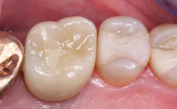
Open contact areas (figure 3)
Why would zirconia contact areas be looser than those from the analog generation? Many tooth preparations are scanned in the mouth rather than making an elastomer impression. Teeth scanned in the mouth without the slight pressure placed on them with a conventional impression in a tray or a double-arch impression are not moved at all when making an impression. Usual tooth mobility from 20 to 200 microns can make a partially open, loose contact area.
The solution: Discuss this challenge with your lab technician, and request that the contact areas need to have approximately 25 microns of extra contour. This is a setting change easily accomplished on the milling machine.
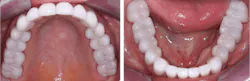
Color match (figure 4)
Full-strength 3Y zirconia is relatively opaque, and usually a thin thickness of ceramic is fired over this type of zirconia to provide optimum shade matching. The feldspathic ceramic used for this purpose wears opposing teeth until it wears off (which takes a few months to several years). At that time, the 3Y zirconia does not wear the opposing tooth enamel, but the zirconia’s true color is exposed.
Other types of zirconia such as 4-5Y (esthetic zirconia) have more optimum color, but their strength is less, and most still have a thin layer of feldspathic ceramic fired over their surface. When it wears off this type of zirconia, it wears opposing teeth.
The solution: Internally staining the zirconia in the green state provides color stability and can be finished and polished by the lab technician, eliminating the need for the feldspathic surface layer.
The future
New forms of zirconia are being developed to help overcome the previously described challenges and others. Zirconia is here to stay. Zirconia has high strength, ease of use, and low cost of fabrication, which make it a nearly optimum crown and fixed prosthesis material. Be patient; improvements are coming.
Summary
Zirconia is continuing to mature as a valid restorative material with millions of crowns now in service, but there are still some challenges to be overcome. This article identifies some of them and makes suggestions about how to overcome them.
Additional reading: Zirconia vs. lithium disilicate
Editor's note: This article appeared in the October 2026 print edition of Dental Economics magazine. Dentists in North America are eligible for a complimentary print subscription. Sign up here.
About the Author
 Gordon J. Christensen, DDS, PhD, MSD
Gordon J. Christensen, DDS, PhD, MSD
Gordon J. Christensen, DDS, PhD, MSD, is founder and CEO of Practical Clinical Courses and cofounder of Clinicians Report. His wife, Rella Christensen, PhD, is the cofounder. PCC is an international dental continuing education organization founded in 1981. Dr. Christensen is a practicing prosthodontist in Provo, Utah.