Bringing the future into focus
by Suzanne Gilman, DDS, FAGD
Dr. Rachel Bella was excited. One of the oral surgeons in her town was offering cone beam or 3-D imaging to dentists and their patients. She was thinking about her patient Jane, a 30-year-old accountant who was having low-grade, chronic pain around her maxillary centrals and laterals for several months. Conventional 2-D periapical X-rays and diagnostic tests did not clarify which tooth, if any, would need root canal therapy. Jane's physician thought that the diagnosis was a sinusitis due to an infection or an allergy, but treatment for those symptoms never gave Jane complete relief. It had been frustrating.
At her next appointment, Dr. Bella explained to Jane that a 3-D scan would show not only the teeth from the front like a regular X-ray, but a side view "slice" in between the teeth. Dr. Bella was pleasantly surprised when the 3-D scan revealed an area on tooth No. 9, and, as a benefit to the endodontist, an unusual curve was also discovered. Jane had root canal therapy on that tooth, and she was happy when the chronic pain went away.
Periapical X-rays have always been, and still are, the choice to diagnose caries and periodontal disease. When viewing bony structures, however, the 2-D limitations of periapicals and panorexes have been a source of uncertainty. It can be difficult to confidently detect the extent of impacted teeth, anomalies, and pathologies. The information needed to completely plan surgeries and implants is restricted.
Until about 10 years ago, we could get some answers from a medical CT scan, in which a fan-shaped X-ray beam creates many image "slices." These 3-D X-ray "pictures" introduce a sagittal plane, in addition to the coronal and axial planes of conventional 2-D X-rays.
Since 2001, instead of sending patients for a medical CT scan, 3-D cone beam computed tomography (CBCT) has been available to dentists for use in their offices. CBCT uses a cone-shaped X-ray beam, is more convenient with less radiation to the patient, and the procedure is similar to taking a panorex. A medical CT scan is 1,200 to 3,300 microsieverts, and the CBCT scan is 24 to 120 microsieverts. (A digital pan is 5 to 15 microsieverts and daily background radiation is 8 microsieverts.)
Depending on the unit, CBCT scan time can be five to 40 seconds, and the reconstruction time - how long it takes the images to appear on the computer screen - is one to six minutes. It takes only a few seconds to find the "slice" you want to study. The patient consult can take place at the same appointment.
Since CBCT does expose the patient to considerable radiation and some added expense, it should only be used as an adjunct, not as a screening tool. But it is solid gold for diagnosing pathology and treatment planning surgery, implants, and orthodontics. In addition, CBCT could well become the standard of care for some of these procedures.
Dr. Lloyd Zivian's patient, 45-year-old Neal, had unusual symptoms on tooth No. 8. The periapical showed a small, distal radiolucency. With only 2-D X-rays to work with, Dr. Zivian would have had to do exploratory surgery, most likely sacrificing the buccal plate. The sagittal view of Neal's CBCT scan showed that the entire facial aspect of the root had resorbed, and the tooth was nonrestorable. Not only did the scan show the extent of the pathological area, it also showed that the buccal plate was intact - excellent prognosis for an implant. Since Neal and Dr. Zivian had a better idea what to expect prior to surgery, Neal was not as fearful consenting to a shorter and more productive surgery.
Patients do not like nasty surprises. A CBCT scan gives both the dentist and patient more information up front. The dentist won't have to backpedal on an already costly treatment plan, and the patient can make a more informed decision.
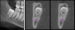
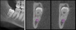
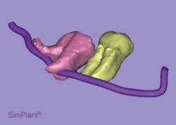
Twenty-five-year-old Joel, a patient of Dr. Mary Frances, presented with a pathological third molar - "superimposed" onto the inferior alveolar nerve, according to the panorex. The CBCT revealed that the roots were actually encircling the nerve, and surgery could be planned with that in mind. Dr. Frances could more realistically discuss the risks and benefits of treatment (or no treatment) with Joel.
CBCT can show the complete extent of an abscess or anomaly, such as a mesiodens, in precise relation to critical anatomy. Surgeries can be shorter and less invasive. CBCT can be used in conjunction with the lab to fabricate drill guides and surgical stents, predicting better esthetic results.
"Three-dimensional radiography should be used in implant placement - it allows a margin of safety that can only benefit the patient," says Dr. Mark McCaffery of Madison, N.J., who uses the i-CAT® in his office. "Being able to do virtual implant planning via SimPlant® or similar programs allows a level of comfort in knowing that there will be few, if any, surprises at the time of tissue reflection and placement."
Terms and concepts
If you are thinking about purchasing a CBCT unit for your office, here are some terms and concepts to consider:
FOV (field of view) - The scans cover different size areas, ranging from 5" x 5" to 14" x 14" or larger, depending on need. Be aware that just as with conventional X-rays, dentists are liable to recognize and inform the patient of any pathology on the scan, not just the area of interest. The larger the FOV, the more anatomy you will be responsible to recognize. It is strongly suggested that you partner with another clinician who is trained to read CBCTs if you are uncertain.
Implant case planning third-party software systems, such as SimPlant®, hire oral maxillofacial radiologists to give an opinion on the scans, and the fee is reasonable. DICOM is the industry standard to transfer radiographic information between computers.
Single Modality Units may have only one FOV setting, therefore giving "extra" radiation to the patient. You take the same scan on every patient and choose the slices you need. Dual Modality Units can have more choices of FOV, along with a separate digital panorex option. Some units have cephalometric capability.
Installation considerations - network, spatial, electrical, and mechanical. If you have an old panorex unit in your office, where is it located? In an alcove or hallway? Chances are you will need more room for the CBCT unit. The dimensions, or footprint, of the i-CAT®, Gendex GXCB-500™, and Vatech Pax-duo3D are about 50" wide, 50" deep, and 70" to 96" high.
You may need to move a wall or devote an entire operatory to the unit. Mike Wendling, service technician at Sullivan-Schein in Pine Brook, N.J., says, "Dentists looking to add a CBCT unit should always be thinking about the space - such as having enough room to take ceph images or for wheelchair access."
A separate, 120-volt dedicated line that serves only the CBCT unit should be installed. The electrician will also have to run the network cables, or leave provisions for the CBCT installer to run the network cables from the unit to the computer. For efficiency in diagnosis and treatment planning, it's recommended to have one computer located close to the unit, to be used only for CBCT image acquisition.
The units mentioned weigh between 500 and 600 pounds, but are freestanding and do not require the walls to be reinforced for extra support.
Call your town municipal department for instructions on how to dispose of the old panorex unit. It can usually go in the trash except for the tubehead. Ask the DEP in your state for the specifics.
If you need help planning for your new unit, representatives from Benco Dental and Vatech will come to your office free of charge to do a site survey before you buy. When you shop for the different units, you will hear words like windows, leveling, beam hardening, scatter, resolution, and voxel. (A voxel is a 3-D pixel). They all come together in varying qualities to form the image.
Look at the image and let that be the determining factor. Benco Dental at CenterPoint has a showroom in Pittston, Pa., displaying several brands of CBCT units, complete with radiography dummies. You can test drive all of the units and find the one that works best for you (www.benco.com/CenterPoint/).
Henry Schein, Benco, and Patterson have a committed interest in providing post-purchase service and support, even if you do not buy your supplies from them. Ask about software updates, compatibility, and universality. Don't let your supplier or your practice-management/chairside software limit the choices of which CBCT unit you can purchase. Do your homework.
Dr. Sharon Gayle was moving her practice to a new location. Her rep was a friend of her brother's, and she had purchased all of her supplies from him for years. She was happy with her chairside software, but it limited her choices of CBCT units. The unit she wanted to buy was not sold by the rep's company. Anticipating the hassle of changing the software annoyed her. She would do it, however, if she didn't have to "settle" for a different CBCT unit.
Her rep told her that there were ways to "bridge" systems and suggested she do research on the Internet and at the convention ... to explore and expand her options. After some research, she found that she was able to integrate her currrent software with the CBCT unit of her choice. Dr. Gayle is happy with both systems and believes she is giving her patients the best care that she can.
Do your research diligently, because the rep won't always think about what is best for you. Find a mentor dentist who has a CBCT in his or her office and discuss all of your options. Ask that dentist: If you were to purchase the unit today, what would you do differently?
Consider the space and the software/network. The image is what you will be working with, so be sure you are happy with it. You already give careful thought to your diagnoses and treatment plans; using that same level of attention while incorporating CBCT into your practice will perpetuate the excellent care you strive to give your patients.
Note: The names of the patients and dentists are pseudonyms.
Suzanne Gilman, DDS, FAGD, graduated from New York University College of Dentistry and practiced general dentistry for 12 years. The creator of the In-Office Dental Radiography Program, she has a passion for explaining topics in dentistry, through written and spoken presentations. Contact her at [email protected] or (973) 229-6256.
For more on this topic, go to www.dentaleconomics.com and search using the following key words: cone beam, CBCT, 3-D imaging, X-rays, implants, Dr. Suzanne Gilman.


