Sensors and Sensibility
By Lorne Lavine, DMD
The modern dental practice has experienced a radical change over the past 15 years. Systems that were paper-based for decades have, in the past few years, been replaced by a digital dental record. Practices are moving toward a chartless concept, where there is no longer a need to maintain a paper chart to view important data. The area that has created the greatest amount of "buzz" in the past two years is digital radiography. In this article, we will explore the benefits of digital radiographs and look at the three basic systems available to incorporate this technology into the dental practice.
There are many reasons why dentists should consider the use of digital radiographs. The main advantages of digital X-rays are the following:
1) Reduced radiation — While this might not be a large concern for most dentists, it is a concern for many patients. When digital systems first came out and film was slower, these systems were marketed as having the ability to take radiographs with 70 to 80 percent less radiation. However, with the advent of faster film, the reduction in radiation is not as pronounced, but still in the 30 to 50 percent range on average.
2) No chemicals — There are few dentists who will object to this! The lack of a need for chemicals and traditional processors (in most digital systems) will translate into fewer lost films, no jammed processors, no need for MSDS sheets, etc.
3) Co-diagnosis — One of the largest hurdles to treatment acceptance is the fact that most patients are unaware of their dental problems. By placing a large image of an X-ray on a monitor that the patient can see, we can begin to include the patient in the diagnosis. Most practitioners who are using digital radiography have reported an increase in patient treatment plan acceptance because of this. While often overlooked, considerable effort should be taken to choose a monitor that has the ideal contrast ratio and other specifications necessary to show diagnostic-quality radiographs.
4) Speed — Depending on the type of system used to digitize radiographs, X-ray images can appear on the computer screen instantaneously, or up to a few minutes later. In all cases, there is a significant time-saving — consider the time it normally takes to load the film into the processor, develop it, mount it, and return it to the doctor. While there are currently two main types of digital systems (more on this shortly), dentists who require immediate images, during procedures such as implant surgery and endodontic treatment, will find the three to five seconds required to view an image from a direct sensor to be a huge benefit to their practice.
5) Image manipulation — One of the biggest advantages of digital images is the ability to modify the image. Adjustments to brightness and contrast, colorization, 3-D, embossing, reversing, and other techniques can be used to assist the dentist in diagnosing dental problems.
6) Electronic submission to insurance carriers and others — Attaching a digital image to an insurance claim can often speed up the process of claim payments. While there is currently no standard in the industry for digital image attachment, I believe this will eventually become commonplace in dentistry. Many dentists also find that the ability to send images through e-mail to referring offices can greatly enhance the level of communication between practitioners.
The options
There are currently three major methods of incorporating digital radiographs into the practice. Each system has advantages and disadvantages, so there is no one system that is "best" for everyone. The three systems that are currently being used are direct sensors, phosphor plates, and digital scanners.
• Direct sensors
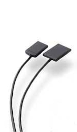
These systems are the most popular digital radiography systems and are the ones that most dentists will see when visiting vendor booths at dental meetings. Some of the major system vendors are Schick Technologies, Kodak, Progeny, Dentrix, and Dent-X. These systems utilize either CCD (Charge-Coupled Device) or CMOS (Complimentary Metal Oxide Semiconductor) sensors that are hooked up directly to the computer in the operatory. Most use a USB connection, although a few still require an internal card.
The upside
• The major advantage to these systems is speed. The sensor will capture the image and send it directly to the computer; images will appear almost instantly in most cases.
• Most systems have their own software for image sorting and manipulation, although the dentists can also use a third-party software that does this, such as XRayVision or Adstra. These image-management programs also integrate quite easily with most of the major practice-management software systems.
• Many of the newer systems have very good image quality, and many dentists consider the image quality to be equal to or better than film.
The downside
• First and foremost for most dentists is cost. The basic systems with a couple of sensors can cost upwards of $20,000, and up to $2,000 for each additional operatory. Most will only come with a one-year warranty, and extended warranties can add thousands of dollars to the initial cost.
• While sensors are not as fragile as they once were, you still need to be very cautious when handling them.
• The sensors are quite a bit thicker than traditional film; the average sensor is about 5 mm thick. Each sensor will have a thin cable attached to the end, which requires some practice in placing correctly in the patient's mouth. Some patients find them to be difficult to tolerate and, of course, they cannot be used with traditional X-ray holders such as the Rinn XCP kits.
• There must be a computer in each operatory where you plan to use the systems.
• There is a bit of a learning curve to negotiate.
• Phosphor plates
These systems are different from the direct sensors in many ways. The system utilizes sensors that are very similar in size and thickness to X-ray film; some are even thinner. The most popular systems include the Scan-X, Orex, and Denoptix. The individual films are taken like a traditional X-ray, then the film is placed either through slots (Scan-X) or in a drum that can hold a full mouth series worth of films. The drum is placed in a scanner and the films will be seen on the computer screen between 90 seconds and four minutes later, depending on the number of films being "processed." The sensors can be reused many times by simply exposing them to a view box, and each sensor costs only $25-$40 to replace.
The upside
• Patients can better tolerate the thinner sensors.
• The system can be set up like a traditional processor, but one that can be used by all the different operatories in the office.
• It is far cheaper to replace one of the sensors.
The downside
• The cost is around $20,000, and most offices can only afford one unit. Therefore, like a traditional processor, if multiple people need to use the unit, there may be a need to wait for the person ahead of you to finish.
• Another issue is that the system must be connected to a computer, so a computer must often be dedicated for this purpose.
• At standard settings, the phosphor plates offer a lower amount of resolution than the direct sensors, although most dentists will probably find that either choice is quite acceptable.
• Direct scan
For the dentist on a budget, and for the dentist who wishes to digitize existing radiographs, this option offers a relatively inexpensive method of incorporating digital radiographs into the dental practice. It involves the use of a scanner with a transparency adapter. The X-rays are placed on the scanner and then scanned; the resulting images can then be seen on the computer monitor. In most cases, the process is made easier by using special software that can organize and manipulate the images. Direct scanning is currently the only system available for digitizing existing X-rays.
The upside
• The cost savings from the other systems can be quite large; a typical scanner and transparency adapter will cost about $1,200, and the software is usually quite reasonable as well.
• This system is ideal for dentists who simply want to archive their X-rays or for the office that wants to save on costs associated with duplicating X-rays.
The downside
• Of course, the quality of the image will be directly related to the quality of the scanner, and in some cases, will be inferior to both the direct sensors and the phosphor plates.
As dentists move into the 21st century, we are faced with an ever-increasing list of choices that will allow us to add technology to the dental practice. While there are only three basic choices available for digital X-ray systems, there are often numerous factors that must be considered when making a purchase. Cost, image quality, space limitations, speed, and a host of other factors must be considered by the dentist before making such a large investment, but it is a necessary exercise if the dentist wants to avoid making a large financial mistake.

