Digital Documentation
Seeing the big picture for guided co-diagnosis
If you’re serious about doing more cosmetic dentistry, remember that it cannot be performed one tooth at a time. Rather, you - and your patients - must address conditions in the esthetic zone in their entirety through what I refer to as guided co-diagnosis. Simply defined, this is the process of garnering the patient’s active participation in the diagnostic phase of their treatment by visually demonstrating their current condition, asking questions, and then proposing the possible treatment outcomes that can achieve their goals.
For purposes of this article, the esthetic zone is defined as the maxillary and mandibular incisors, cuspids, and premolars - the very basis of cosmetic dentistry. To engage patients in effective guided co-diagnosis, you first must be able to capture and visually display their complete current condition. Then, you must engage their participation in the diagnostic process by asking questions. Finally, once you understand their perspective and clinical condition, you guide them through the cosmetic dental possibilities for enhancing their smile and appearance.
Why photographic documentation?
Time and time again, it’s been said that a picture is worth a thousand words. When it comes to understanding dental conditions and proposed treatments, an image can say it all. In fact, patients often understand what they see better than what they hear. The dynamics of the human face during conversation and smiling can often hide disharmonies that become readily visible when observing static digital images of the face and closeup views of the teeth. Therefore, you should not be without a great digital camera, a closeup lens system, and a laptop computer with PowerPoint. When combined, these resources will enable you to digitally document the patient’s cosmetic condition and successfully guide him or her through the co-diagnosis process.
With your narration of the digital images, a patient will be able to make educated choices about accepting proposed care. The limitations of single-tooth restorative dentistry can be more clearly explained and understood, and your patient is less likely to make a choice to repair just one tooth. In this regard, patient education is of paramount importance in increasing the amount of cosmetic dentistry you perform.
Currently, much dentistry is defined by dental insurance policy coverage. If a patient only receives $1,000 of coverage per year, usually only single-tooth restorations are planned because the patient cannot “see” the big picture.
Although this type of covered care may address the immediate need for repairing a broken tooth, if the treatment is required in the esthetic zone, the repair may continue to be a visible reminder of the failure of communication. This leads to restorative dentistry that is clearly indicated, but not necessarily cosmetic. That is why incorporating guided co-diagnosis into the practice routine can become a profitable exercise for both the patient and dentist.
Why ask questions?
The questions you will ask your patient will help lead you understand what makes a disharmonious smile. From there, the natural progression of the consultation turns to the restorative steps that can be taken to correct these disharmonies. Some sample questions to ask may include:
✦ What do you like about your smile?
✦ Would you like your teeth to look whiter?
✦ Are you happy with the appearance of your teeth?
✦ What would you change if you could?
✦ What don’t you like about your smile?
✦ Do you like the arrangement of your teeth?
There are literally hundreds of questions that you could ask to guide a patient through the process of co-diagnosis. Each question sets the stage for elevating the patient’s overall understanding of the principles of smile design.
Digitally document
Co-diagnosis of the esthetic zone requires that patients be able to see what you see so that they can discuss what they like and what they don’t like. You, as the clinician, must also be able to visually identify what is clinically necessary from a restorative standpoint.
I take digital photographs of every new patient who enters my practice. This is usually a series of six views that clearly demonstrate the condition of the teeth in the esthetic zone. Along with a full series of radiographs and mounted study casts (when indicated), these digital images are the foundation of the consultation - or guided co-diagnosis - appointment. In essence, they guide the discussion with patients regarding their feelings about their smile and what they would like to change.
These images are incorporated into a PowerPoint presentation that becomes the road map for the treatment plan discussed with the patient. During this discussion, not only is the patient’s current oral condition addressed and visually displayed, but computer generated simulations of the possible results of proposed treatments are also presented.
Capturing digital photographic images serves another purpose. It keeps you focused on the cosmetic aspects of dentistry. For example, when performing an initial examination without digital photographs, I believe it is quite possible to focus on individual teeth and adopt more of a “repair-the-damaged-tooth” philosophy.
When you see the entire esthetic zone, all of the cosmetic and restorative possibilities - and even necessities - become collectively apparent. Specifically, when you view the digital images with the patient, pay special attention to identifying some of the following examples of disharmonies, based on smile design principles.
Tooth from disharmonies
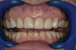
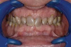
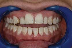
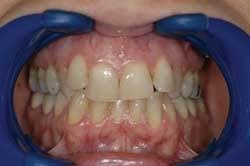
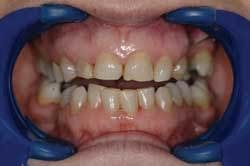
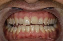
The shape of the central incisors can make or break a great smile. Some of the problems that are readily apparent on digital images (Figs. 1 and 2) include:
✱ Height-to-width ratio of the central incisors
✱ Height of the central incisors
✱ Incisal embrasure form
✱ Balance and symmetry
✱ Proportions of the teeth/repeating ratios
null
Tooth color discrepancies
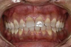
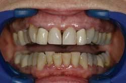
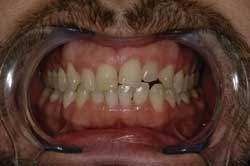
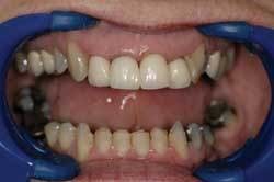
Stated another way, digital images help dentists (as clinicians) as well as patients see the bigger picture. For example, with a digital image of multiple teeth shown side-by-side, it is possible to identify discrepancies with tooth color. This includes color variations that have resulted from crowns being placed at different times, discolored composite or alloy restorations, or problems with discolored tooth structure (Figs. 3 and 4, next page).
The digital image often will illustrate a mosaic of color. Allowing the patient to observe the image and be a part of co-diagnosis often leads to questions about how the smile - or, more specifically, the color disharmony - can be improved. Solving the patient’s color disharmony with multiunit restorative dentistry is a great example of the cosmetic rejuvenation that becomes possible through the use of guided co-diagnosis.
But remember that cosmetic dentistry cannot be performed one restoration at a time. Pointing out to the patient how these color problems occur in the first place is a great justification and rationale for explaining why cosmetic treatments are planned the way they are - with multiple, appropriately selected restorations seated simultaneously to create a harmonious, functional smile.
Failing restorative treatments
Perhaps a discrepancy in tooth color was an obvious example. Digital documentation of the patient’s smile is also beneficial in emphasizing the detrimental effects to his or her appearance caused by failing restorative dentistry. These effects could include black margins on old crowns, recurrent decay on failing composites, or stained enamel in which teeth have been chipped (Figs. 5 and 6).
Without digital images, the tendency often is to repair, replace, or restore the failing restorations without regard to the overall appearance of the surrounding teeth. This can further create the appearance of a mosaic of color throughout the esthetic zone.
Problems with tooth position
Tooth position disharmonies also can be overlooked if the practitioner is simply looking for problems with either hard or soft tissue. One of the biggest deficiencies in terms of tooth position that can be identified through an analysis of digital images is the lack of fullness in the premolar areas (Figs. 7 and 8). A deficiency of this kind gives the patient the appearance of having very prominent front teeth and dark corridors in the posterior region.
Some other types of tooth position problems include midline shift, axial inclinations of the teeth, and arch form. Crowding and excess space also are problems that become more easily recognizable when viewing digital images. Patients gain a new perspective on the symmetry - or lack thereof - of their smiles, and are more receptive to the many corrective measures available.
Problems with the soft tissue
The condition and appearance of the soft tissue often can create disharmonies in the smile that are more easily identifiable on digital images (Figs. 9 and 10). Some of the most common types of soft-tissue problems include:
• Gingival height symmetry
• Zenith placement
• Gingival embrasure form
Conclusion
Once you incorporate digital images of the esthetic zone, you will begin to see many types of cosmetic disharmonies. By asking simple questions, you then will be able to guide patients through a co-diagnosis process that will help them better understand the need for multiunit restorative care (Figs. 11 and 12). Smile design will become second nature, and you will be surprised at how frank patients can be when discussing their smiles. This is an inevitable progression. As you incorporate guided co-diagnosis, you (as a clinician) begin to see the bigger picture, and are less likely to compromise a diagnosis for the sake of tooth-by-tooth appointments and procedures. With greater multiunit restorative case acceptance, you begin to see that the benefits of smile design are not only a beautiful smile, but also better function and a healthier periodontium.
The process of guided co-diagnosis enables patients to understand the need for multiunit restorative dentistry to resolve their esthetic disharmony. Treatment plans that involve a combination of porcelain laminate veneers, crowns, and bridges then become the only realistic measures to correct crowding, mosaics of color, or excess space. Thus, your patients can see and understand that a great cosmetic result cannot be achieved “one tooth at a time.”
References available upon request.
Dr. Harry A. Long is the 2004 winner of the Cosmetic Practice of the Year Award. He is also the 1973 recipient of the Victor O. Lucia Award for proficiency in prosthetic dentistry. A clinical instructor for the Aesthetic Advantage dental continuum, he is a member of the peer review committee for the Passaic County Dental Society. Dr. Long can be contacted by telephone at (973) 694-5101, or via e-mail at [email protected].


