Methacrylate-functionalized calcium phosphate: A clinical primer
Feb. 28, 2022
4 min read
Creating a long-term seal of a restorative material to dentin has long been a significant clinical challenge. This deficiency leads to marginal breakdown, recurrent decay, and ultimately the replacement of the restoration with further loss of tooth structure. Materials with methacrylate-functionalized calcium phosphate (MCP)
technology are helping to solve the problem of insufficient long-term sealing of enamel and dentin. MCP aids in the transfer of key ions for remineralization and the building of apatite crystals. More specifically, when placed in a dental composite, MCP facilitates the biointeractive transfer of calcium, phosphate, and fluoride from the material and the oral environment to promote the formation of hydroxyapatite bridges between tooth structure and restorative material. In the process, MCP also facilitates elevated esthetics.Additional reading:
- Dental bonding: Technique matters
- Combining glass ionomer, IDS, and CEREC for intelligent hybrid restorations
Case no. 1
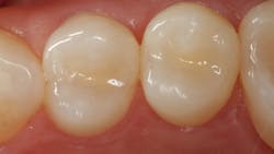
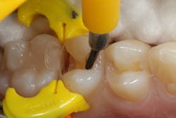
Caries lesions were detected in the central grooves of nos. 12 and 13 using the caries detection mode of a SoproCare intraoral camera (Acteon). After preparation was completed (figure 1) using a fissurotomy bur (SS White), the dentin andenamel were etched using 38% phosphoric acid (Etch-Rite, Pulpdent) for 15 seconds and then thoroughly rinsed and dried. The preparation was then treated with an antimicrobial cavity cleanser (FiteBac, FiteBac Dental), which improved adhesive capability and became part of the restorative-dentin interface. Next, an adhesive bonding agent (Dentastic Uno-Duo, Pulpdent) was applied to the cavity surface. It is important to note that the bonding agent will not inhibit the exchange of ions from the restorative material.
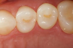
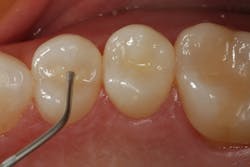
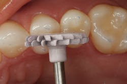
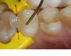
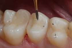
Activa Presto with Crysta MCP technology (Pulpdent) was selected as a restorative material. The composite was dispensed into the cavity preparation (figure 2) and then light-cured. A narrow, “bullet-type” 20-fluted finishing carbide (SS White) was used to make occlusal adjustments and accentuate anatomic detail as required. Final polish was accomplished using A.S.A.P composite polishers (Clinician’s Choice; figure 3).
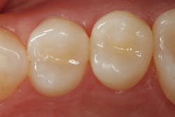
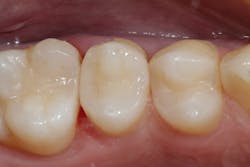
Crysta MCP technology may facilitate the formation of apatite crystals at the tooth-restorative interface to maintain the integrity of the margin and help prevent recurrent decay or staining. Figure 4 shows the completed restoration.
Case no. 2
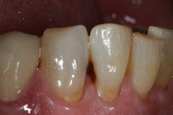
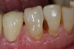
Figure 5 is a preoperative facial view of a class III caries lesion on the distoproximal aspect of no. 26. This lesion is mostly all on the root surface. Any lesion whose preparation extends onto a root surface may suffer a compromisedmarginal seal when traditional composite is used as the restorative material due to difficulty of isolation. In this case, there was also a class III caries lesion on the mesial surface of no. 27.Figure 6 shows the removal of caries and completion of the preparation. Note that a diode laser was used to make the gingival margin of the cavity preparation supragingival to facilitate a good gingival seal to the margin with the MCP-containing restorative material.
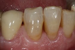
The completed restorations are shown from a facial aspect in figure 7. The highly esthetic match using Activa Presto shade A6 makes it impossible to find the restoration in the tooth.
Case no. 3
Use of Crysta MCP technology is an excellent choice for conservative class II restorations. A sectional matrix system is recommended for class II placement to limit the material to the preparation and avoid proximal excess. Activa Presto was layered in 2 mm increments into the cavity preparation(figures 8 and 9). MCP technology will help combat the top cause of class II composite failure—lack of seal of the gingival margin of the proximal box of the cavity preparation. This is the area where most recurrent decay begins and results in ultimate failure. Figure 10 shows minor occlusal adjustment using the small “bullet-shaped” finishing carbide (FG-7901, SS White). Figure 11 shows an occlusal view of the completed restoration.
Case no. 4
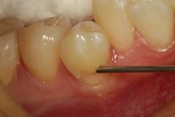
Class V caries lesions often involve the root surface on the facial aspect at and below the gingival crest. These areas are particularly hard to successfully seal with conventional composite materials. Figure 12 shows a class V preparationbeing filled with Activa Presto. Note the gingival margin of the preparation is slightly supragingival on the root surface. MCP technology will help form a scaffold (nucleation site)
for deposition of calcium and phosphate ions, forming and maintaining an “apatite-like” seal of this area at the gingival margin. If the patient is not fastidious with home-care regimens, MCP may help maintain the
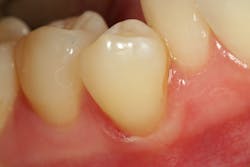
integrity of the tooth-restorative interface. Figure 13 shows the completed restoration.
Editor's note: This article originally appeared in the February 2022 print edition of Dental Economics.
About the Author

Robert A. Lowe, DDS, FACD, FADI, FAGD, FASDA, FICD
Robert A. Lowe, DDS, FACD, FADI, FAGD, FASDA, FICD, is a cosmetic dentist and associate professor in the department of reconstructive and rehabilitative sciences at the Medical University of South Carolina James B. Edwards College of Dental Medicine. He works as an educator, author, and consultant for several dental manufacturers. Dr. Lowe is a recipient of the Gordon Christensen Outstanding Lecturer Award.
Sign up for our eNewsletters
Get the latest news and updates
