Choosing between complete (crown) or partial coverage (inlay and onlay) restorations: What will last?
There is significant increase in life expectancy and in the number of retained natural teeth at an older age.1,2 Although preventive dentistry is widely practiced, dental caries remains a prevalent oral disease, causing irreversible loss of tooth structure. Tooth structure loss through attrition, abrasion, erosion, or combination of these is also persisting among the world population.3-5
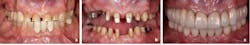
In a very simplified sense, restorative dentistry aims to remove decayed dental tissues and restore what is lost with a material to replicate its shape, shade, and function. Based on the amount of remaining tooth structure, a clinician can choose between complete (crown) or a partial coverage (inlay or onlay) restoration.1,6,7
Preparations for complete coverage restorations are more invasive,8 and because the tooth can have large failing restorations and decay, there is widespread opinion that complete coverage restorations result in higher number of loss of tooth vitality.8 Thus, over many years, restorative dentistry has favored minimally invasive procedures to preserve as much tooth structure as possible. Thus modern dentistry is shifting toward partial coverage restorations even in the teeth that require lingual, occlusal, and buccal coverage restorations.9,10 Partial coverage restorations are indeed more conservative due to the nature of the preparation and the path of insertion.8
Additional reading: When should teeth be removed?
Teeth have become a sign of social status, and patients request tooth-colored restorations. This drive for more natural-looking materials has led the industry to move away from gold and other precious metals. In fact, a dentist is faced with many resin and ceramic products, making it difficult to decide which material to choose for a clinical situation.11
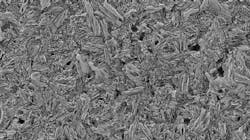
Lithium disilicate was introduced to the dental market in the early 2000s as IPS e.max Press (Ivoclar Vivadent) and has become a popular material for anterior restorations, combining excellent esthetics with acceptable mechanical properties.12 However, its flexural strength of 470 MPa and fracture toughness of 2.54 MPa have led to questioning the use of lithium disilicate restorations in the posterior region,13 where occlusal loads are higher14 and where materials with higher flexural strength and fracture toughness such as monolithic zirconia have been preferred.15,16
Moreover, there is a widespread assumption that e.max lithium disilicate glass ceramic with a thickness less than 1 mm is more susceptible to catastrophic fracture,17 which leads to more invasive tooth preparations or avoidance of the material.
Which type of glass ceramic restoration in the posterior dentition, complete or partial, performs better over a long time remains unanswered. There is also no evidence-based answer for the question of which type of restoration, complete or partial, results in higher incidence in the need for endodontic therapy.
Therefore, we performed a prospective clinical study with twofold aim:
- To compare long-term clinical survival and the clinical factors influencing the outcomes of adhesively bonded e.max lithium disilicate glass ceramic complete and partial coverage restorations, and to evaluate the performance of e.max lithium disilicate glass ceramic restorations in the posterior teeth.
- To assess the incidence of teeth requiring endodontic therapy after receiving either a complete or a partial coverage glass ceramic restoration with up to 36 years of follow-up.
The prospective study was initiated in 1985 and the database parameters as well as the recall method were adopted from previously published studies of the same group of researchers.18-25 Clinical confounding variables evaluated were: dental arch, tooth position in the dental arch, age and sex of participant, ceramic thickness, and type of restoration.
We are sharing our scientific findings to help clinicians in decision-making and provide evidence-based answers when choosing restorations and materials.
The experimental method
Participants in this study were at least 20 years of age and had demonstrated full-mouth plaque score (FMPS) and full-mouth bleeding score (FMBS) 25%. Teeth included in the study had adequate periodontal support; no or limited mobility; and adequate remaining tooth structure for the choice of a single-unit, defect-specific, partial or complete coverage restoration; and had to be vital.
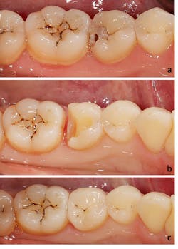
The decision as to which type of glass ceramic restoration (complete or partial coverage) considered the extent of damage, presence of fracture lines, and resistance and retention form.6,7
For partial coverage restorations, defect-specific tooth preparations removed all the caries and created proper retention form. Inlay or onlay partial coverage preparation design was then chosen based on the remaining tooth structure.1 The complete coverage restorations were approximately 1.2 mm in depth, and marginal finishing burs were employed.
Restorations were completed in a conventional manner utilizing medium body polyether (Impregum, 3M ESPE) impression material. Lost-wax technique and a glass ceramic pressing system were then used to fabricate the definitive restorations.
The restorations
After clinical evaluation and necessary adjustment, all restorations were etched (4.5% buffered hydrofluoric acid, IPS Ceramic Etching Gel; Ivoclar Vivadent) for 20 seconds, and silane (Monobond Plus; Ivoclar Vivadent) was applied for 60 seconds. The teeth were etched with 38% phosphoric acid (Etch-Rite; Pulpdent), coated with a desensitizer (Gluma Desensitizer; Kulzer), and dentin bonded (Excite; Ivoclar Vivadent). The restorations were adhesively luted with a light-polymerizing resin (Variolink II; Ivoclar Vivadent) activated with an LED polymerization light (Bluephase Style; Ivoclar Vivadent). All the excess cement was removed thoroughly.
Prior to cementation, the following parameters were entered or determined: type of ceramics, type of restoration, restoration thickness measured by calipers at up to seven points (mesial, distal, buccal, lingual, mesial-occlusal, midocclusal, distal-occlusal), tooth position, age and sex of the patient. The restorations with at least one of the above-described measurement points less than 1 mm were grouped in the thickness of less than 1 mm.
The participants were routinely recalled every six months. The status of the restoration(s) was evaluated, and the incidence of postprosthetic root canal therapy was assessed.
The results
Cumulative survival
The clinical performance of 6,683 units up to 36 years was excellent, with the estimated cumulative survival of 96.35%.There were 84 biological failures (defined as tooth needing postprosthetic endodontic therapy) recorded, out of which 61 occurred in posterior complete, 12 in posterior partial, and 11 in anterior complete coverage restorations, providing a crude estimate of an annual percentage of biological failures of 0.16% with the survivor function time at 35.6 years. The incidence of 84 endodontic therapies occurred during a cumulative monitoring period of 51,564 years, with an overall survival rate of 96.35%.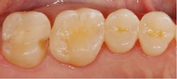
Posterior vs. anterior
Posterior complete coverage restorations had statistically significant higher biological failure rate than anterior. The overall clinical performance of posterior complete coverage restorations relative to biological failure was still high with a cumulative survival of 95.15% over 35 years.
Posterior complete vs. partial coverage
There was no difference in biological failure rate between posterior complete and partial coverage restorations. First and second molars had the highest rate of postprosthetic endodontic therapy in both arches.
Failure rate by sex
Failure rate by age
There was no difference in biological failure rate of different age groups.
Time to failure
The survival of 2,392 posterior e.max lithium disilicate complete and partial coverage restorations placed in 738 participants was evaluated at 17 years. Only 22 failures were recorded with a 16-year cumulative survival of 96%.
The average time to failure was 3.5 years. No debonded restorations were recorded. The majority of failures (77%) occurred within 6 years. There were no failures beyond 8 years of service.
Type of bonding
The data indicated that acid etched and adhesively bonded monolithic IPS e.max pressed lithium disilicate complete (97%) and partial (95%) coverage restorations exhibited excellent survival in the posterior teeth.
Complete vs. partial
No statistically significant difference was found between complete and partial coverage restorations.
Clinicians widely use complete coverage restorations, especially in the posterior dentition.26 However, the opinion leaders in the dental community are becoming more critical of the preparation protocols needed for these restorations.27,28 Quantification of preparation types showed a 68% to 76% removal of tooth structure for complete coverage restorations, which is significantly more than the amount removed for partial coverage restorations.8
Other concerns associated with crown preparation were postprosthetic need for endodontic therapy, weakening of the tooth, catastrophic root fracture, and finally, the need for extraction.7,29 In contrast, indirect partial coverage restorations can offer a minimally invasive treatment procedure with reliable occlusal schemes.30
Although the difference was not statistically significant, neither in survival rates nor in the need of endodontic therapy, it provides scientific evidence for encouraging clinicians to use partial coverage restorations in the posterior teeth given that remaining tooth structure is adequate. This is opposed to always leaning toward complete coverage restorations, which, in some respect, is still considered to be a gold standard among clinicians.
However, this should not serve as an encouragement for fabricating partial coverage restorations where they’re not indicated.
The newer dental trend entails insertion of two restorations (buccal and lingual) for a single tooth. Although, some studies have been published using this technique, their number of patients and units is very small and follow-up time is shorter.9,10
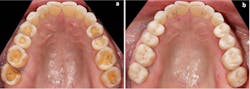
If a tooth requires buccal, occlusal, and lingual coverage restorations, it is more convenient and financially feasible to fabricate single complete coverage restoration. Because there is no difference in the survival or loss of tooth vitality, complete coverage restorations should not be neglected. For a tooth that has significant loss of tooth structure, or requires restoration of several surfaces due to severe wear, a crown is a well-validated treatment modality.
The role of materials
Ceramic materials have evolved dramatically over the last two decades,11 with so many ceramic materials that choice is based on personal preference and opinion, rather than evidence. One such widely spread opinion-based assumption is to avoid the use of lithium disilicate in posterior dentition due to high occlusal loads that could lead to premature fractures. In 2,392 posterior restorations studied over 17 years, only 22 fractures were recorded. Most failures occurred within the first 6 years and then declined, with five additional failures at 8 years. There were no additional failures in the 236 restorations with time in service of from 8 to 17 years. This declining failure rate suggests a lack of fatigue degradation in these longer-term restorations and will be explored in a future publication.
Of the 22 failures, 18 occurred in the molar region, and no debonding was seen. Our group has previously reported that Dicor glass ceramic has a higher risk of fracture in the molar region. Although there is also a trend of higher failure in the molar region for the lithium disilicate, no statistical significance was yielded. Even for the mandibular second molars that showed the highest failure rate (six failures), the estimated annual risk of failure was only 0.4% and without statistical significance. This provides evidence for choosing lithium disilicate for the molar region.
Influence of thickness
Thickness of ceramic material had no influence on the incidence of biologic failures in posterior complete, posterior partial, and anterior complete coverage restorations. Restorations with surfaces less than 1 mm and greater than or equal to 1 mm performed similarly over 17 years.
Another widely spread but clinically unsupported opinion is to avoid using lithium disilicate with thickness greater than 1 mm.31,32 This recommendation translates into removing additional tooth structure to create the desired greater than or equal to1 mm clearance.
In the current study, the variable of thickness had no effect on the survival of complete and partial coverage restorations in the posterior teeth. The restorations with at least one surface with a thickness less than 1 mm performed similarly to those with a thickness of 1 mm or more. Similar findings have also been reported in clinical and in vitro studies.
The lack of influence of the restoration thickness can be explained by the adhesive luting protocol used for both complete and partial coverage restorations in the present study. It is well established that the mechanical properties of ceramics increase with adhesion, which explains the findings in a series of studies.20,27,28,33,34
These findings should encourage clinicians to be less invasive during preparation and minimize the clearance required for e.max lithium disilicate glass ceramic restorations to save as much of the tooth structure as possible.
Influence of covariates
Covariates such as tooth position, sex, and age demonstrated no effect on survival.
Both age and sex are considered confounding variables in medical and dental studies, as they entail factors such as occlusal force, oral hygiene, and diet.35
In our studies, the assessment of age and sex as confounding variables was completed, and no significant effect on survival was recorded. This provides scientific and clinical evidence for choosing lithium disilicate glass ceramic complete and partial coverage restorations in male and female patients regardless of age.
Conclusion
Lithium disilicate is an etchable glass ceramic, and a strong micromechanical bonding to tooth structure is developed,11 resulting in improved physical properties of the restoration.36,37 In the present study, all the restorations were adhesively luted by using the dentin bonding agent followed by the adhesive cement Variolink. This may explain the higher overall survival rate of over 96% and the fact that no restoration debonded over this observation time.
The low rate of loss of tooth vitality may be attributed to the adhesive cementation protocol used. Phosphoric acid etching of the abutment teeth removes smear layer and bacteria. Dentin sealing using Gluma, followed by a dentin bonding agent and resin cement, results in a reliable seal that could prevent bacterial leakage and possible contamination and micromotion between the core substrate (dentin and/or enamel) and ceramics.
Each patient has an individual clinical scenario with varying health conditions and remaining tooth structure. Therefore, it is important that restorative dentists make an evidence-based selection of material and treatment method. We hope to have provided evidence-based answers to these everyday clinical questions.
Editor's note: This article appeared in the February 2022 print edition of Dental Economics magazine. Dentists in North America are eligible for a complimentary print subscription. Sign up here.
References
- The glossary of prosthodontic terms: ninth edition. J Prosthet Dent. 2017;117(5S):e1-e105. doi:10.1016/j.prosdent.2016.12.001
- Frencken JE, Sharma P, Stenhouse L, Green D, Laverty D, Dietrich T. Global epidemiology of dental caries and severe periodontitis - a comprehensive review. J Clin Periodontol. 2017;44(Suppl 18):S94-S105. doi:10.1111/jcpe.12677
- de Melo MA, Passos VF, Lima JP, Parente GC, Rodrigues LK, Santiago SL. Erosive potential of processed and fresh orange juice on human enamel. J Dent Child (Chic). 2015;82(1):10-15.
- Hattab FN, Yassin OM. Etiology and diagnosis of tooth wear: a literature review and presentation of selected cases. Int J Prosthodont. 2000;13(2):101-107.
- Maher RL, Hanlon J, Hajjar ER. Clinical consequences of polypharmacy in elderly. Expert Opin Drug Saf. 2014;13(1):57-65.
- Goodacre CJ, Campagni WV, Aquilino SA. Tooth preparations for complete crowns: an art form based on scientific principles. J Prosthet Dent. 2001;85(4):363-376.
- Goodacre CJ, Spolnik KJ. The prosthodontic management of endodontically treated teeth: a literature review. Part I. Success and failure data, treatment concepts. J Prosthodont. 1994;3(4):243-250.
- Edelhoff D, Sorensen JA. Tooth structure removal associated with various preparation designs for posterior teeth. Int J Periodontics Restorative Dent. 2002;22(3):241-249.
- Mainjot AKJ. The one-step no-prep technique: a straightforward and minimally invasive approach for full-mouth rehabilitation of worn dentition using polymer-infiltrated ceramic network (PICN) CAD-CAM prostheses. J Esthet Restor Dent. 2020;32(2):141-149.
- Oudkerk J, Eldafrawy M, Bekaert S, Grenade C, Vanheusden A, Mainjot A. The one-step no-prep approach for full-mouth rehabilitation of worn dentition using PICN CAD-CAM restorations: 2-yr results of a prospective clinical study. J Dent. 2020;92:103245. doi:10.1016/j.jdent.2019.103245
- Gracis S, Thompson VP, Ferencz JL, Silva NR, Bonfante EA. A new classification system for all-ceramic and ceramic-like restorative materials. Int J Prosthodont. 2015;28(3):227-235.
- Willard A, Gabriel Chu TM. The science and application of IPS e.Max dental ceramic. Kaohsiung J Med Sci. 2018;34(4):238-242.
- Alkadi L, Ruse ND. Fracture toughness of two lithium disilicate dental glass ceramics. J Prosthet Dent. 2016;116(4):591-596.
- Manns A, Rojas V, Van Diest N, Rojas D, Sobral C. Comparative study of molar and incisor bite forces regarding deciduous, mixed, and definitive dentition. Cranio. 2020:1-8.
- Blatz MB, Vonderheide M, Conejo J. The effect of resin bonding on long-term success of high-strength ceramics. J Dent Res. 2018;97(2):132-139.
- Ortorp A, Kihl ML, Carlsson GE. A 5-year retrospective study of survival of zirconia single crowns fitted in a private clinical setting. J Dent. 2012;40(6):527-530.
- Shahmoradi M, Wan B, Zhang Z, Wilson T, Swain M, Li Q. Monolithic crowns fracture analysis: The effect of material properties, cusp angle and crown thickness. Dent Mater. 2020;36(8):1038-1051.
- Malament KA, Margvelashvili-Malament M, Natto ZS, Thompson V, Rekow D, Att W. 10.9-year survival of pressed acid etched monolithic e.max lithium disilicate glass-ceramic partial coverage restorations: performance and outcomes as a function of tooth position, age, sex, and the type of partial coverage restoration (inlay or onlay). J Prosthet Dent. 2021;126(4):523-532. doi:10.1016/j.prosdent.2020.07.015
- Malament KA, Margvelashvili-Malament M, Natto ZS, Thompson V, Rekow D, Att W. Comparison of 16.9-year survival of pressed acid etched e.max lithium disilicate glass-ceramic complete and partial coverage restorations in posterior teeth: performance and outcomes as a function of tooth position, age, sex, and thickness of ceramic material. J Prosthet Dent. 2021;126(4):533-545. doi:10.1016/j.prosdent.2020.08.013
- Malament KA, Natto ZS, Thompson V, Rekow D, Eckert S, Weber HP. Ten-year survival of pressed, acid-etched e.max lithium disilicate monolithic and bilayered complete-coverage restorations: performance and outcomes as a function of tooth position and age. J Prosthet Dent. 2019;121(5):782-790.
doi:10.1016/j.prosdent.2018.11.024 - Malament KA, Socransky SS. Survival of Dicor glass-ceramic dental restorations over 14 years: part II. Effect of thickness of Dicor material and design of tooth preparation. J Prosthet Dent. 1999;81(6):662-667.
- Malament KA, Socransky SS. Survival of Dicor glass-ceramic dental restorations over 14 years: part I. Survival of Dicor complete coverage restorations and effect of internal surface acid etching, tooth position, gender, and age. J Prosthet Dent. 1999;81(1):23-32.
- Malament KA, Socransky SS. Survival of Dicor glass-ceramic dental restorations over 16 years: part III. Effect of luting agent and tooth or tooth-substitute core structure. J Prosthet Dent. 2001;86(5):511-519.
- Malament KA, Socransky SS. Survival of Dicor glass-ceramic dental restorations over 20 years: part IV. The effects of combinations of variables. Int J Prosthodont. 2010;23(2):134-140.
- Malament KA, Socransky SS, Thompson V, Rekow D. Survival of glass-ceramic materials and involved clinical risk: variables affecting long-term survival. Pract Proced Aesthet Dent. 2003;(Suppl):5-11.
- Pjetursson BE, Lang NP. Prosthetic treatment planning on the basis of scientific evidence. J Oral Rehabil. 2008;35(Suppl 1):72-79.
- Edelhoff D, Guth JF, Erdelt K, Brix O, Liebermann A. Clinical performance of occlusal onlays made of lithium disilicate ceramic in patients with severe tooth wear up to 11 years. Dent Mater. 2019;35(9):1319-1330.
- Guess PC, Selz CF, Steinhart YN, Stampf S, Strub JR. Prospective clinical split-mouth study of pressed and CAD/CAM all-ceramic partial-coverage restorations: 7-year results. Int J Prosthodont. 2013;26(1):21-25.
- Goodacre CJ, Bernal G, Rungcharassaeng K, Kan JY. Clinical complications in fixed prosthodontics. J Prosthet Dent. 2003;90(1):31-41.
- Opdam N, Frankenberger R, Magne P. From ‘direct versus indirect’ toward an integrated restorative concept in the posterior dentition. Oper Dent. 2016;41(S7):S27-S34.
- Guess PC, Schultheis S, Wolkewitz M, Zhang Y, Strub JR. Influence of preparation design and ceramic thicknesses on fracture resistance and failure modes of premolar partial coverage restorations. J Prosthet Dent. 2013;110(4):264-273.
- Nordahl N, Vult von Steyern P, Larsson C. Fracture strength of ceramic monolithic crown systems of different thickness. J Oral Sci. 2015;57(3):255-261.
- Bakeman EM, Rego N, Chaiyabutr Y, Kois JC. Influence of ceramic thickness and ceramic materials on fracture resistance of posterior partial coverage restorations. Oper Dent. 2015;40(2):211-217.
- Guess PC, Zavanelli RA, Silva NR, Bonfante EA, Coelho PG, Thompson VP. Monolithic CAD/CAM lithium disilicate versus veneered Y-TZP crowns: comparison of failure modes and reliability after fatigue. Int J Prosthodont. 2010;23(5):434-442.
- Richards D, Clarkson J. Evidence-based Dentistry: Managing Information for Better Practice. Quintessence Publishing; 2008:80-82.
- Bailey LF, Bennett RJ. DICOR surface treatments for enhanced bonding. J Dent Res. 1988;67(6):925-931.
- Pagniano RP, Seghi RR, Rosenstiel SF, Wang R, Katsube N. The effect of a layer of resin luting agent on the biaxial flexure strength of two all-ceramic systems. J Prosthet Dent. 2005;93(5):459-466.
Editor’s note: For more details on this study, access the full-length paper at dentaleconomics.com/malament.
About the Author
Kenneth A. Malament DDS, MScD
Kenneth A. Malament DDS, MScD, received his DDS from NYU College of Dentistry and a specialty certificate and master’s degree from Boston University School of Graduate Dentistry. Dr. Malament has a full time practice limited to prosthodontics in Boston that includes a dental laboratory with master dental technologists. A Past-President of the American Board of Prosthodontics, he is a clinical professor at Tufts University and a course director in the postgraduate department of prosthodontics.

Mariam Margvelashvili-Malament, DMD, PhD, MSc
Mariam Margvelashvili-Malament, DMD, PhD, MSc is an assistant professor in the department of prosthodontics at Tufts University School of Dental Medicine. She is a diplomate of the American Board of Prosthodontics and fellow of American College of Prosthodontics. Dr. Margvelashvili-Malament received her DMD from the Tbilisi State University in Georgia in 2007, also master of science and doctoral degrees in dental materials from the University of Siena, Italy, in 2009 and 2013, respectively.