Ask Dr. Christensen
by Gordon J. Christensen, DDS, MSD, PhD
In this monthly feature, Dr. Gordon Christensen addresses the most frequently asked questions from Dental Economics® readers. If you would like to submit a question to Dr. Christensen, please send an e-mail to [email protected].
For more on this topic, go to www.dentaleconomics.com and search using the following key words: radiographs, diagnostic standards, radiation, digital vs. analog radiographs, Dr. Gordon J. Christensen.
Q What is considered to be the standard for diagnostic oral radiographs in a typical general practice? I have heard and observed everything from only a panoramic radiograph to the conventional 14 to 18 exposure full-mouth series. I want to offer my patients good service for my exams, but I do not want to overdo the radiation exposure. What do you suggest?A Your question is more complex than it appears. There are numerous types of radiographs available today, and they relate to the specific needs of patients. I will discuss your question from various aspects in an attempt to make it more practical and understandable.Digital vs. analog radiographs
The dental profession in North America has been relatively slow to implement digital radiography compared to other developed countries. The advantages of digital radiography are well known, and there are significant reasons to accept this major change as an important improvement in diagnosis and treatment- planning. The advantages are:
- Immediate observation of images
- Magnifying images
- Measuring distances between various features on radiographs
- Enhancing images with texture or color
- toring images for immediate retrieval
- Great for patient education
- Increasing or decreasing contrast is easy
- Elimination of the developer
- Easy to e-mail to patients and other practitioners
- Lower radiation than analog
However, there are a few disadvantages including the following:
- Cost is significant
- Conversion from conventional to digital requires time and effort
- Learning to use the concept takes time
- Breakage or loss of sensors
- Thickness of sensors
- Rigidity of sensors
- Current lack of universal use of digital
In the event that you have not integrated digital radiography into your practice, I suggest you do so as soon as you can afford to purchase the devices. In a few years, analog radiography will vanish, as has film for photographs.
Most dentists who have digital radiography use CCD/MOS systems that allow for immediate observation of the images; however, some are using phosphor plates that are less expensive and have thinner sensors. Phosphor plates do not provide an immediate image, and you must place the plate into a processor to allow an image to appear on your monitor.
Type of practice and usual radiographic needs
The type of patients usually treated in a specific practice relates directly to the radiographs considered to be most appropriate for diagnosis and treatment-planning. I will use actual patient radiographs to demonstrate the relative acceptability of the various types of current digital radiographs.
Periapical radiographs: Most dentists use periapical radiographs daily for a variety of diagnostic and documentary reasons. I suggest having the availability of a state-of-the-art periapical digital radiographic device. Some of the most popular brands are: Schick, DEXIS®, Kodak, Sirona, GE, Gendex, and many others.
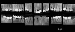
Full-mouth periapical radiographic series (Fig. 1): When many teeth are in the mouth, most dentists prefer making periapical radiographs of all of the teeth. A typical full-mouth series includes the following separate images: molars (4), premolars (4), canines (4), anterior teeth (2), for a total of 14 separate images. These 14 images are usually augmented with four bitewing radiographs. If many teeth are missing, only a few periapical radiographs are usually indicated. If only a few teeth are remaining in the mouth, a panoramic radiograph may be adequate. The ability of either analog (D-speed film) or digital radiographs to show initial dental caries has been studied and compared. Neither type of radiograph shows initial caries well. Additionally, panoramic radiographs do not show initial caries. Some dentists have eliminated the 14-to-18 image, full-mouth series and use only panoramic radiographs. Patient education using only periapical radiographs is difficult, since many teeth are duplicated in the various views, and this is confusing to patients.
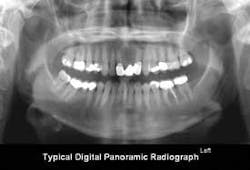
Panoramic radiographs (Fig. 2): The current digital panoramic radiograph devices are excellent, and the radiographs provided by them show an overall, easily understood view of the oral structures. Panoramic radiographs are excellent for initial screening and can provide adequate singular radiographs for some types of patients, especially those with only a few teeth remaining or edentulous patients.
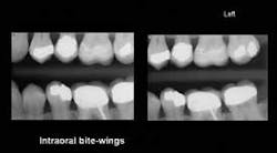
Intraoral bitewing radiographs (Figs. 3A and 3B): These radiographs, used for nearly a century, have allowed observation and diagnosis of initial dental caries in the past. However, with the advent of reduced radiation, bite-wing radiographs have decreased in value relative to showing initial caries. Additionally, when the currently available, relatively thick, sensor holders are used, they often open the mouth so far that the cervical areas of the teeth are not clearly visible on the radiograph. When using the intraoral bite- wings for patient education, the duplication of some teeth can be confusing to patients.
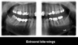
Extraoral bitewing radiographs (Fig. 4): This concept, currently available from PLANMECA USA and Sirona, shows the teeth, the periapical areas, and the related bony structures. These images are very educational for patients, and allow better diagnostic capability for practitioners than intraoral bitewings.
Other radiographs: You asked about radiographs for conventional diagnosis and treatment-planning in general practice. However, as you know, cephalometric, tomographic, cone beam (3D), and other radiographs are also available. They are used for specific needs, including orthodontics, implant placement, detection of oral pathology, and other situations, but usually not for conventional diagnosis and treatment-planning in typical general practices.
What types of radiographs should you use for an initial exam and treatment-planning session? The needs of your practice dictate your radiographic needs. As I observe dental practices around the country, I see the following varied use of radiographs:
1) No radiographs. Believe it or not, some dentists avoid using radiographs as much as possible. I do not suggest this approach.
2) Panoramic radiographs only. Although this concept shows the major tooth positioning, large carious lesions, and overall jaw anatomy, these radiographs often lack detail adequate to show specific tooth pathology. However, they often are adequate for removable partial or complete denture diagnosis and treatment-planning.
3) Panoramic radiographs and bitewing radiographs. A significant percentage of dentists use this concept. For many patients, it is probably acceptable. I suggest that if a practitioner is using just a panoramic radiograph and bitewings, periapical radiographs should be used for suspect teeth.
4) A full-mouth series of 14-to-18 separate periapical images usually includes one or two bitewings on each side. This concept satisfies the diagnostic needs of many general dentists, periodontists, and others; however, because of the duplication of teeth on the images, it has limitations relative to educating patients.
5) Option 4 accompanied with a panoramic radiograph is a concept used by some dentists. This option provides a significant amount of radiation and may be excessive, but for patients with many teeth remaining and numerous oral problems, it offers the best ability for dentist diagnosis and treatment-planning and for patient education.
Your decision on what type of radiographic services you will provide should be based on personal choice and what's best for your practice and your patients. I do not have one protocol for my practice. The need for radiographs varies from patient to patient.
Our new video, “Maxillofacial Digital Radiography — Simplified,” shows how to take routinely used digital radiographs easily and with predictability. It is excellent for educating staff on how to take digital radiographs.
For more information, contact Practical Clinical Courses at (800) 223-6569, or visit thh PCC Web site at www.pccdental.com.
Dr. Christensen is a practicing prosthodontist in Provo, Utah, and Dean of the Scottsdale Center for Dentistry. He is the founder and director of Practical Clinical Courses, an international continuing-education organization initiated in 1981 for dental professionals. Dr. Christensen is a cofounder (with his wife, Rella) and senior consultant of CLINICIANS REPORT (formerly Clinical Research Associates), which since 1976 has conducted research in dentistry.


