Cone beam technology: Clinical applications
by Matthew R. Lark, DDS, MAGD, FICD, FACD
A brief overview of the many software applications available for the CBCT.
For more on this topic, go to www.dentaleconomics.com and search using the following key words: computerized axial tomography, CAT scanners, Cone Beam Computerized Tomography, Hounsfield units, orthogonal reconstruction.
Cone Beam Computerized Tomography (CBCT) is the most diverse emerging technology to reach dentistry in decades. Clinical applications touch every aspect of dentistry, from general practice to orthodontics, periodontics, implantology, prosthodontics, oral maxillofacial surgery, endodontics, orofacial pain, sleep dentistry, and oral medicine practice. This wide range of applications to dental disciplines has changed dentistry forever into a three-dimensional world. We no longer have to interpolate the third dimension from arcane measurements of stone dental casts and two-dimensional radiographs. Why guess when we can know?
Applications of the CBCT depend upon innovative software that compiles the DICOM data from the CBCT scan and reconstructs it into usable images which can be studied, measured, and virtually manipulated for surgical treatment-planning.
Reading the CBCT
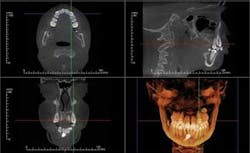
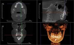
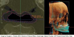
All CBCT manufacturers include OEM (Original Equipment Manufacturer) software with their machines. This software varies with the specific units, but generally includes a method of viewing the scan in an interactive frontal, sagittal, and coronal mode known as Multi-Planer Reconstruction (MPR). Each scan is reviewed first by browsing through the cross sections in all three planes and noting any findings, with particular attention paid to the diagnostic condition of interest (Figure 1).
Specialized orthogonal cross sections, such as panoramic planings, are performed to elicit a more directed view of areas of interest. The Hounsfield density measures the relative density of the involved structures and offers insight into the nature of the lesion. Most OEM software includes a 3-D viewer which can be applied to the scan to observe the volume of the target and its juxtaposition to other structures. Measuring tools in the software enable precise measurements in both the 2-D and 3-D images. The 3-D images can be cropped to decrease superimposition of adjacent structures — surface rendered to view skin and airway volume — all of which can be rotated for viewing at any angles. Most OEM packages have a function which displays corrected tomographic AP and coronal slices through the temporomandibular joints.
Doctors who have in-office CBCT scanners will have access to all OEM software. However, the doctor referring scans to outside facilities will also require access to the OEM software. Some machines, such as I-CAT or GENDEX, provide I-Cat Vision for referrals to view. The scan can be sent via e-mail or disc to the referring doctor for interpretation. Some CBCT scanners do not provide the off-site viewing option, so the practitioner will have to:
Therefore, you need to know whether the OEM software is available to the referring doctors before purchasing a CBCT unit if you plan to offer a scanning service.
The CBCT scans can be helpful in tracking infection using the OEM 2-D and 3-D software. Some accessory canals and fractures can be visualized with high resolution scans. The endodontist can use the scans to determine the origin of an infectious tract. Sinus infections, cysts, and polyps are easily visualized in the MPR views. You also can observe the Atlanto-Occipital Junction and several cervical vertebrae for pathology. Bony defects — which look benign in the 2-D preview — often become remarkable when viewed in 3-D.
Most specialized software applications are after-market products or services sold separately for the scanning machines. The decision to purchase software depends on the needs of the practice. Categories of software include: 1) orthodontic planning, 2) oral maxillo-facial surgery planning, 3) implant planning, and 4) TMD diagnostic planning.
Orthodontic planning
For decades, the dental stone casts and cephalometric analysis have been the cornerstone of orthodontic treatmentplanning. With the introduction of CAD/CAM, orthodontists were able to apply computer technology to manipulate the arch forms and tooth positions. Facial soft tissue, jointbased occlusion, and dento-skeletal information were interpolated from photographs or mounted casts. CBCT orthodontic software gives the diagnostician all of the information required to treatment-plan, track, and archive the complex orthodontic case.
Dolphin Imaging® (dolphinimaging.com) and InVivoDental by Anatomage® (www.anatomage.com) are two software developers who are on the cutting edge of software for orthodontic and orthognathic treatment-planning and archiving. Both packages offer conventional cephalometric analysis based upon digitized 3-D volume renderings. The soft tissue and facial photos (in 2-D or stereographic formats via 3-D MD camera system) can be superimposed and phased in over the skeleton with a translucency feature. This feature enables integrated dento-facial analysis showing how the soft tissues are influenced by dento-skeletal positions. Location, size, and density of lesions and bone cysts — as well as positions impacted and supernumerary teeth — can be rendered in 2-D and 3-D to help determine the best approach for surgical access. (Figure 2).
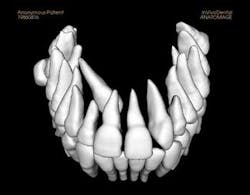
CBCT images can be modeled by Anatomage in the traditional form of digital study casts. They can then be viewed and morphed into desired tooth positions. The pre/post treatment CBCT scans can be superimposed showing the positional change between the two, which is actually 4D imaging (Figure 3). An animated, digital morph movie can be made to show the before and after morphing.
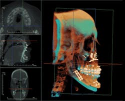
Anatomage is at the forefront of this "Dynamic CBCT" with its ability to superimpose two CBCT scans to visualize actual changes over time or projected changes with modeling simulations (Figure 4).
Research in 3-D cephalometric data will some day superannuate the 2-D cephalometric analyses. CBCT adds the dimension of joint-based occlusion (Piper) to the analysis. This encompasses the influence of condylar morphology and position, intra-articular pathology, growth disturbances, and retrograde remodeling on the functional mandibular positions.
3-D measurements will allow insights into the influence of anterior-constricted envelope (Kois) relative condylar position in the genesis of temporomandibular joint disorders, unstable anterior tooth position, constrictive anterior wear patterning, and observing how latent mandibular growth can account for distalized condyles and trapped anterior teeth. In observing Class II dentitions and anterior open bites, the relationship of the decreased ramus height, condylar growth lag, degenerative disease, or anterior displaced discs can illicit the cause of an occlusal shift toward a Class II dentition (Class III shifts to Class I).
Maxillofacial surgery studies
The Anatomage software employs various methods to filter out metallic scatter, producing a highly selective, clean 3-D volume in which structures can be easily selected and isolated for analysis. This process of clean isolation permits views of teeth, airway, temporomandibular condyles, the mandibular complex, and sinuses. Furthermore, it allows volumetric measurement and positional manipulation to formulate surgical or prosthetic projections.
Any structure that can be imaged can be rendered as a 3-D stereolithic model, which can be manufactured with SLA 3D printers. This capability allows surgeons to pre-plan surgeries such as joint replacement surgeries or major facial reconstructions on tabletop models. Bone plates can be preformed, implants selected, templates made for graft sizing, hands-on manipulation done for mock surgery, and arch bar and surgical stents fabricated.
Sleep dentistry airway imaging
The emerging field of sleep dentistry will benefit from the ability to indirectly measure the airway volume and record the areas of maximum airway constriction. Anatomage has a feature which isolates the airway and allows 3-D study of the influence of mandibular position or spinal encroachment on airway volume. With this study, a dentist can determine whether a sleep apnea appliance will improve the airway status with a George Gauge positional CBCT scan. The two scans with and without the mandibular advancement device show the exact change in the airway, allowing for a treatment efficacy study (Figure 5).
Temporomandibular joint studies
Temporomandibular studies can be formed from a combination of the OEM software and the Anatomage software. There are six important temporomandibular joint images required to analyze TM disorders.
- Axial corrected sagittal slices
- Axial corrected coronal slices
- Ramus height measurements
- Condyle to fossa size ratio
- Condylar axial surface area in square mm
- 3-D volume renderings of skull and condylar isolation
The temporomandibular joint images can be viewed relative to the occlusal position; thus, if there is a change in the status of the occlusion, the pre/post images can be taken and superimposed in the 4-D volume rendered format. In this way, changes in temporomandibular joint position as a result of occlusal change, or vice versa, can be determined. This application can elucidate controversies regarding condylar position relative to occlusal position, and demonstrate how changes in one element affect the other. For example, this imaging can help explain how a change in condylar height or position may cause previously unexplained changes in the occlusion.
Implant dentistry software applications
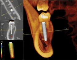
There are many software programs available for CBCT assisted implant surgery. Virtual Implant Placement® (VIP by Implantlogic, Implantlogic.com) and SimPlant (Materialize) are two programs that provide the ability to treatment-plan cases from prosthetically driven scanning appliances, to implant selection and placement, to stealth surgical guide-assisted implant placement. Implant software packages work with most popular implant systems, allowing the selection of the exact implant required, thus reducing the need to overstock expensive implant inventory. There are other proprietary systems which offer CBCT implant planning. The best software is the one that functions for the dentist and the implant system that works best in her/his hands.
Guided-implant placement planning precisely determines the margins of error from vital structures, such as the mandibular nerve, lingual concavities, floor of the nose, facial undercuts, adjacent roots, and the maxillary sinus. CBCT helps surgeons operate within the "zone of safety" and has significantly increased the predictability of 3-D implant placement (Figure 6).
Stealth prosthetically driven implant surgery treatment-planning<
Prosthetically-driven stealth implant surgery represents the future of implant dentistry. Using CBCT, the exact tooth positional requirements are determined early in the treatment-planning process. "People don't want implants, they want teeth," so placing the implant exactly in the desired position supports better implant/prosthesis mechanics, thus improving the functional and esthetic results.
Preliminary scan and prosthetic diagnostic wax-up
A preliminary scan determines whether adequate bone volume and quality exist to properly support the planned implant-borne prosthesis. The bone volume is assessed in 3-D, and length, height, and width characteristics are planned according to the prosthetic requirement. Bone augmentation procedures are carried out when inadequate bone quality or morphology are found.
Treatment-planning with VIP software
The Implantlogic.com Web site contains an excellent set of movie tutorials on the entire stealth-guided implant surgery process. This software can be used with implant inventory from over 20 notable implant companies.
A CT scanning appliance is fabricated on a stone cast where the desired replacement tooth positions are determined using a mounted, diagnostic wax-up on a semi-adjustable articulator. Next, the implant support teeth are transformed into barium-sulfate impregnated acrylic-trial teeth and processed into the acrylic-scanning guide. In the future, this step will be completed with a virtual diagnostic wax-up using 3-D metallic fiducial makers. They will be placed using a jig to allow 3-D loci for realigning the scanning appliance to the CBCT image. Next, the scanning appliance is placed in the mouth, and the patient is scanned with the desired arch parallel to the cone-beam sensor.
The CBCT scan is uploaded to the planning software and the final implant positions and selections are determined according to sound treatment-planning protocol. Implants are planned 1.5 mm from adjacent teeth and 3 mm from adjacent implants. This helps maintain healthy support bone and papilla adjacent to the implants. The mandibular nerves, adjacent tooth roots, sinuses, and other structures are determined and the case is planned within the desired zone of safety. The implant plan is e-mailed to the laboratory and the scanning appliance is sent back for processing. The laboratory technician and consulting dental surgeon evaluate the plan, and final corrections are made prior to surgical-guide fabrication.
The surgery guide, which contains a 3-D drill-position sleeve, is now used to drill the surgical osteotomy. This procedure often can be performed in a stealth manner, meaning through a minimal tissue punch instead of a surgical flap. Stealth surgery decreases postoperative complications, since the periosteal and gingival blood supply remain intact and mental nerve dissection is not necessary. However, it is important to make sure that bone and soft-tissue properties meet the implant support and esthetic requirements prior to using the stealth approach. Otherwise, conventional flap designs may be used. In single-stage implants, the perimucosal healing cap is placed and the tooth is restored after osseous integration is complete.
Learning which CBCT imaging applications are right
This article is a brief overview of the multitude of software applications available for the CBCT. There are many other software companies and applications on the market which could not be covered in this short space. Each year, a 3-D symposium is held to discuss all of the new applications of CBCT. The symposium features hardware and software developers, as well as vendors displaying all of the new applications for CBCT. The symposium also includes talks by dentists and dental radiologists discussing the recent research and applications in CBCT technology. Information can be found at [email protected].
When making a decision to purchase CBCT application software, ask the vendor if a time-limited trial is available to see if it performs the tasks you desire. Some software programs are very complicated to operate, and some are very simple. It is important to discuss your practice needs and your computer skills for effective software utilization. Ask if you will incur additional charges for maintenance and upgrades. Also ask if there is free, limited-function shareware for your referring doctors.
As more dentists use CBCT, the unleashing of the scientific imagination has led to the development of software applications that were unfathomable five years ago. Patients are benefiting from the advent of CBCT by receiving better diagnostics, enhanced surgical and orthodontic treatment-planning, and ultimately, safer and more predictable surgeries in the areas of implantology, orthodontics, maxillo-facial reconstructive trauma, and temporomandibular joint disorders. This also facilitates patient education, understanding, and treatment acceptance.
CBCT has created a paradigm shift for the dental profession. Development of new CBCT technological applications for clinical dentistry and research will guide us toward innovative solutions for tomorrow's patients. The time has come for dentists to emerge from their dip tanks and embrace the new era of 3-D dental radiology with CBCT.
Matthew R. Lark, DDS, MAGD, FICD, FACD, has practiced general dentistry in Toledo, Ohio, for the past 26 years. He is the current president of the American Academy of Orofacial Pain. He is also an assistant clinical professor at the University of Toledo Medical Center, Department of Dentistry. You may contact Dr. Lark by e-mail at [email protected].
