Livin' large: special effects of digital X-ray
by Lorin Berland, DDS
For more on this topic, go to www.dentaleconomics.com and search using the following key words: digital radiography, digital technology, magnification, digital X-rays.
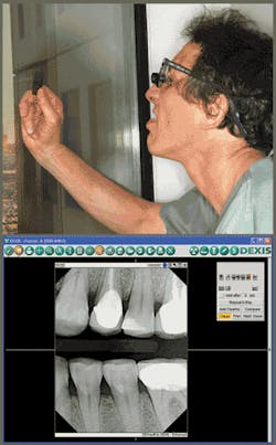
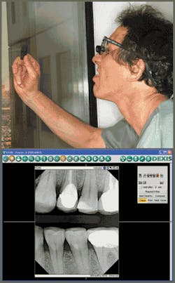
Whoever thinks "Good things come in small packages" is obviously not a dentist ... and especially does not spend time squinting at postage-stamp-sized film X-rays all day. For diagnosis and treatment planning, our office mantra has become "Bigger is better."
Dentists need magnification for every aspect of the patient's treatment, including diagnosis. By the end of dental school, most graduates probably will own a pair of loupes and may even enlist the aid of a microscope for some procedures.
While magnification tools are imperative for directly examining the oral complex, digital radiography's image enhancement capabilities have clearly improved imaging and, as a result, diagnosis and treatment planning. I haven't used film X-rays for 10 years, and I stopped using film panos more than a year ago. But I still remember the hassles and limitations with traditional radiography and know that my job has been made easier thanks to digital technology.
Here is what I have learned about improving diagnosis with digital technology:
• Digital X-rays are vastly superior to film because they can be zoomed in on or enlarged. The magnification, clarity, and detail of digital radiography cannot compare to traditional film. Frequently patients arrive in my office and can't figure out which tooth is hurting. With a digital X-ray, I have the capacity to expand and magnify images and detect cracks, decay, or periapical radiolucencies that I might miss on film.
• Digital X-rays are not just larger; they also offer enhancement opportunities. Using enhancements, I have the added ability to alter the brightness or contrast to detect the smallest flaws. My DEXIS® digital system has an application called ClearVu™ that uses mathematical algorithms to enhance details for better detection of tooth fractures or caries. With traditional film, I was dependent on the chemistry of the developing materials to give me a clear X-ray. Unless the processor was primed, at the right temperature, ready and perfect, with fresh developing and fixing solution, the picture quality was inconsistent. Also, film X-rays diminish with time and handling. Digital images are consistent and easy to read, each time you open them on the screen.
Another command, "Invert," flips the black and white areas, much like a photographic negative. When compared to the noninverted image, and toggled back and forth, this function can help figure the position of canals within the root of the tooth and verify the presence of caries or abscesses.
• Flexible display options create effective patient presentation and consultations. Diagnosis is not important unless it's co-diagnosis; the patient and doctor have to be on the same "screen." Additional contrasts offer options besides just enlarging the image. Certain enhancement functions, such as "Darker" and "Relief," are especially helpful when viewing the X-rays with patients. "Darker" mode obviously darkens the image, but it also compensates for varying densities, such as with anterior and posterior teeth. "Relief" mode gives the images a 3-D look (without those funny cardboard glasses used in the movies) to pinpoint small density differences. With film, patients were hard-pressed to see the X-ray, let alone understand any details, so they just couldn't fully understand the facts that help them make informed decisions about their treatment.
With film, about the only thing you can do is flip it over. With digital images and a few clicks of the mouse, the patient's dentition can be larger than life and definitely more defined. It's time to stop squinting at tiny films and start showing patients "the big picture" of their dental options.
Dr. Lorin Berland is an internationally acclaimed cosmetic dentist and one of the most published authorities in the professional dental and general media. Dr. Berland, a Fellow of the American Academy of Cosmetic Dentistry, is the co-creator of the Lorin Library Smile Style Guide, www.denturewearers.com, and is the founder of Arts District Dentistry, a multidoctor specialty practice in Dallas that pioneered the concept of spa dentistry. Dr. Berland was honored by the American Academy of Cosmetic Dentistry with the 2008 Outstanding Contribution to the Art and Science of Cosmetic Dentistry Award.

