A case report on everyday workflows with new Multi-Direct Capture scanning technology: From hygiene to final restoration
What you'll learn in this article
- How Multi-Direct Capture technology in the iTero Lumina enables faster, more photorealistic scans—making it feasible to incorporate full-arch scanning into hygiene and new-patient visits
- Why 3D visualizations during consultations improve patient understanding, boost case acceptance, and enhance communication between providers and patients
- How routine digital scanning supports earlier diagnosis by RDHs and leads to better lab outcomes through improved margin clarity, prep accuracy, and reduced remake rates
Digital scanning technology has been present in dentistry in some form since the 1980s. While digital scanning in dentistry has been mostly focused on restorative dentistry, recent developments in scanning technology increase the applications within the practice. The newest scanning technology, iTero Multi-Direct Capture, brings speed, photorealism, and accuracy that make this technology suitable to use in every aspect of a dental practice.
Most digital scanners available for purchase as of 2024 use confocal imaging to capture a digital model. This technology has been well researched and developed. Using a single camera, a scanner takes images of dental structures, records them, and maps them using distances between known points.1 This technology produces reliable models, but due to the single-image capture, it can only move at a certain speed. Processing speed of the computer can quicken the process, but it is still a single stream of data being captured.
Multi-Direct Capture technology utilizes five different cameras and six light sources to expand the field of view and depth of focus. This increases the speed and smoothness of the scanning experience, as well as providing an astoundingly photorealistic digital model.
I have been using digital scanning technology in my practice since 2009. Initially, we used our digital scanner in the manner that most dentists do—as a replacement for impression material for indirect restorations. Beginning in 2017, we began to change how often we used our scanner.
It started with a patient in one of my hygiene chairs for a recall examination. She had a visible fracture line on the distal of her lower right second molar, and I recommended a crown. I was trying to explain to her why she needed this restoration, but she left her hearing aids at home, so she was having trouble hearing me. A picture is worth a thousand words, right? With this in mind, I got out my DSLR camera and attempted to take a photo. The battery was dead. In haste to get her exam finished and her crown scheduled, I grabbed my iTero Element 2 scanner. We did a quick (four-minute) scan, and I showed her the crack on her tooth. She could immediately see the issue and quickly understood the treatment plan.
This serendipitous event led me to a realization. Patients understand more when they can see what we see, especially in three dimensions. We began scanning every new patient. This was an investment in time, but it paid off. Within three months, our practice production was up nearly 30%. When I talk about this idea with other dental practices, I generally get pushback about that investment in scan time during an already rushed dental appointment. With Multi-Direct Capture technology on our iTero Lumina, our scan time is around 35 seconds per arch. The idea that scan time is a barrier from scanning in our new-patient and recall appointments is a thing of the past thanks to this new technology.
Case study: From recall to treatment
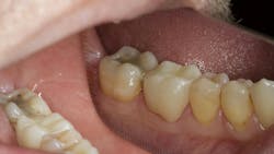
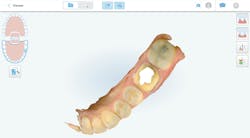
Our patient Chris presented for his regular routine recall visit with one of our dental hygienists (figure 1). When she asked him if he had any issues, Chris reported a slight discomfort on his lower left side when chewing. As part of his normal recall experience, our dental hygienist did a “wellness” scan on Chris using our iTero Lumina scanner.
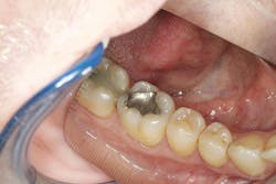
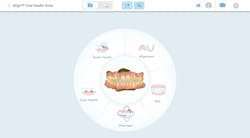
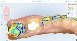
During my examination, I noted a large amalgam restoration on the lower left first molar with an apparent crack down the distal marginal ridge. No percussion tenderness was noted on any area in the quadrant except for the distolingual cusp of the lower left first molar. A bite test also showed tenderness in this area and nowhere else. A cold test indicated that the lower left first molar was vital and had no lingering effects. My treatment plan for Chris was simple: a crown and buildup on his lower left first molar. My dental hygienist had the scan loaded on the screen of our iTero Lumina using the Align Oral Health Suite. Align Oral Health Suite is a tool that helps clinicians educate their patients on all aspects of their dental health using the scanner, including tooth health, gum health, bite, and alignment (figure 2).
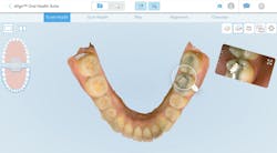
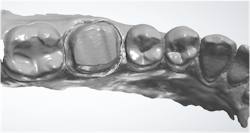
Using Align Oral Health Suite, I was able to show Chris his lower first molar, both on the scan and an isolated intraoral camera photo taken during scanning (figure 3). He was able to easily see the issue with his tooth, quickly agreed to treatment, and provided consent. The lower left molar was prepared for a full-coverage crown. The old amalgam restoration was removed and replaced with a dual-cured, fiber-reinforced opaque buildup. The preparation was refined, and a size 0 cord was packed in the sulcus. The preparation was then imaged using the iTero Lumina scanner.
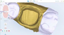
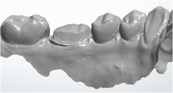
The Multi-Direct Capture technology within the iTero Lumina allowed for fast, accurate scanning of the preparation. Total scan time for the prep, operative quadrant, opposing quadrant, and bite was 77 seconds (figure 4). Using the robust restorative software on the iTero Lumina, I was able to verify adequate occlusal reduction (figure 5), as well as accurately mark the margins for my laboratory technician (figure 6). The prescription was filled out and sent to the laboratory directly from the iTero Lumina, while my assistant fabricated and cemented a provisional crown for the patient.
A dental laboratory perspective
As a robust and busy digital dental laboratory, we handle hundreds of digital scans per day. Our laboratory purposefully focuses on doctors who utilize digital scanning, because we find we get better results with them than those who use analog impression materials. Our internal data shows that remake percentages go down when doctors use digital scanning. In addition, adequate reduction occurs more often, which makes for more successful restorations. We all know that smooth margins, adequate tissue management, and clean, dry preparations are vital to success with digital scans; however, some scans are just easier to work with than others. The iTero Lumina produces scans like this.
What we notice is a brilliantly lifelike image, startlingly different in detail, color rendition, and representation of polychromatic characteristics from other confocal imaging scanners. Lumina’s photorealistic capture of detail sets a new standard for imaging quality and communication (figure 7).
While the definition of the prep and margin is very clear and definable, it is the lifelike rendition of color, textures, and light reflection that gives Lumina scans unequaled clarity and definition. This is important not only for margin detection, but surface details of the preparation and surrounding dentition. The excellent chromatic representation allows for definitive identification of margins against tissue, a feature never before seen in previous scanners (figure 8). Lumina’s remarkable depth-of-field is evidenced by complete interproximal capture (versus missing data) and clear exposure balance, resulting in realistic surface texture and clarity (figure 9).
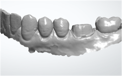
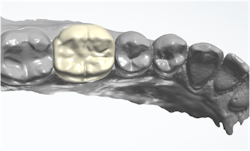
In this case, we were able to design an anatomic, well-contoured crown for Dr. Austin’s patient, Chris, which was milled out of traditional 3Y zirconia dioxide (figures 10 and 11).
The final restoration
Chris returned to the practice for his crown insert five days after the preparation. Cindy’s lab, 38Smiles, focuses on producing high-quality zirconia crowns from digital scans within three days of preparation. Allowing a day or two for shipping, we routinely insert single-unit crowns on day five after preparation. We find this increases the ease of the delivery appointment for both the patient and clinician, as well as decreasing the chances for a remake versus the traditional two weeks for delivery.
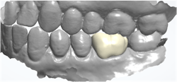
Chris’s provisional was removed, and the preparation was cleaned. The zirconia crown from the lab required no adjustment and was cemented with RMGI luting cement. This visit took a total of nine minutes from start to finish. With the accuracy of the iTero Lumina scanner, and the expeditious nature of 38Smiles, we find this to be a common occurrence in our indirect restorative procedures. The patient has tolerated this procedure well and reported no pain or discomfort after the restoration was placed (figure 12).

A summary
Digital scanning technology is a staple of modern restorative dentistry. Moving from confocal imaging to Multi-Direct Capture offers a number of advantages for indirect restorations, including accuracy, increased margin readability, and photorealistic capture. As restorative dentists, this is what most of us focus on. However, another advantage of scanning with Multi-Direct Capture is the ability to use this technology in the hygiene and wellness visit to help educate and inform our patients of their dental needs. When patients can visualize (in three dimensions with photorealism) what their dental issues are, case acceptance goes up. This can change the trajectory of a dental practice, increasing production, collections, and patient satisfaction. When the financial aspects of a dental practice strengthen, in addition to an increase in patient satisfaction, that will inevitably lead to an increase in dentist satisfaction as well.
Editor's note: This article appeared in the July/August 2025 print edition of Dental Economics magazine. Dentists in North America are eligible for a complimentary print subscription. Sign up here.
Reference
- Richert R, Goujat A, Venet L, et al. Intraoral scanner technologies: a review to make a successful impression. J Healthc Eng. 2017;2017:8427595. doi:10.1155/2017/8427595
About the Author

Joshua Austin, DDS, MAGD
Joshua Austin, DDS, MAGD, is a graduate and former faculty member of the University of Texas Health Science Center at San Antonio School of Dentistry. Author of Dental Economics’ Pearls for Your Practice column, Dr. Austin lectures nationally on products, dental technology, online reputation management, and social media. He maintains a full-time restorative dentistry private practice in San Antonio, Texas. You may contact Dr. Austin at [email protected].
Updated June 21, 2023
Cindy Lam-Vuong, CDT
Co-Owner of 38 Smiles Digital Dental
Cindy Lam-Vuong, CDT, is co-owner of 38 Smiles Digital Dental. She has a BA in communications and specialty certificates in LEAN and LEAD. With 30-plus years of experience, her diverse career includes 25 years as a technician specializing in veneers and smile makeovers. She participated in beta-testing of materials, including the formulations of BruxZir and BruxZir Esthetic, and digital software and intraoral hardware development while acting as production manager and general manager at Glidewell Laboratories.