Better all the way around!
by Jeffrey B. Dalin, DDS, FACD, FAGD, FIGD
Digital radiography represents the cornerstone of the digital diagnosis technologies in my practice. In this series of articles, I will discuss its benefits and applications over and over again. I cannot imagine returning to the use of conventional X-ray film. I am always amazed at the objections I hear from dentists who say they don't believe the time is right to introduce digital radiography into their practices. My advice is clear: The time is right and that time is now!
Dentistry was first introduced to digital radiography in France in the late 1980s. Of course, the systems we use today are far removed from the large, expensive, and unreliable systems that were available then. The technology has evolved in stages and has matured greatly since that time. Affordability, portability, usability, and diagnostic quality all have improved dramatically in the last decade.
Diagnostic image quality speaks to why we all practice dentistry. I remember how the image quality of the early digital systems was greatly inferior to that of X-ray film. Not any more. Technological breakthroughs in sensors and software have resulted in digital images that are at least as good as those of X-ray film. And, due to enlarged image size and enhancement tools, digital images often allow for superior diagnoses to be made.
I could go on, but let me summarize the other benefits of digital radiography:
- less radiation exposure to your patients
- instant on-screen images
- rapid and superior diagnoses
- image-enhancement capabilities (such as magnification, contrast, and coloring)
- improved patient acceptance and education
- total elimination of darkroom, chemicals, and film
- easy image sharing with other dentists or third-party payers
So ... digital radiography is better for your patients and better for your practice!
Now, I want to give you an illustration of why I am such an enthusiastic advocate of digital radiography. I always liked the saying, "A picture is worth a thousand words." Let me explain why.
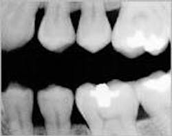
Recently, a patient came in to see me for a routine checkup. He reported some occasional pain in his lower left quadrant. In Figure 1, you will see a reproduction of the digital bitewing that we took in my office that day.
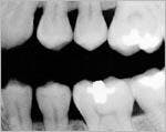
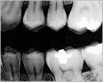
We noticed a vertical line running from the bottom of his amalgam restoration down to the pulp chamber. When we enhanced the image using the Clear-Vu tool in our DEXIS digital radiography system (see Figure 2), the fracture was clearly evident. We then took a conventional X-ray film, shot at regular speed and hand-developed at 68 degrees. By contrast, the vertical line was not evident on that film at all!
Any doubt you might have had about image quality and diagnostic abilities of digital images should be laid to rest after viewing these examples. Digital radiography is here to stay. Your patients and your practice will surely benefit from this exciting technology.
Jeffrey B. Dalin, DDS, FACD, FAGD, FIGD, practices general dentistry in St. Louis. He also is the editor of St. Louis Dentistry Magazine and spokesperson and critical-issue-response-team chairperson for the Greater St. Louis Dental Society. His address on the Internet is www.dfdasmiles.com. Contact him by email at [email protected] by phone at (314) 567-5612, or by fax at (314) 567-9047.


