XRAY Files #1 — A dynamic duo comes to the rescue!
by Terry L. Myers, DDS, FAGD
For more on this topic, go to www.dentaleconomics.com and search using the following key words: digital imaging technology, digital radiology, 3-D imaging, Dr. Terry Myers.
In our busy, stressful lives, many of us would benefit from having our own hero — not the kind who flies with a cape or tangles up the bad guys in a web, but an everyday hero, one who calms a hectic schedule, soothes our nerves, and generally makes life easier. Digital imaging technology often plays the hero in my practice. Convenient and speedy, it rescues us from time-wasting tasks, such as waiting for film X-rays to develop. Plus, latest advancements such as 3-D imaging literally give me detailed “X-ray vision” so my patients understand diagnosis and accept treatment.
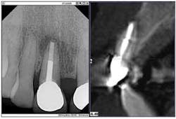
My intraoral X-ray (DEXIS) and cone beam (Gendex GXCB-500) systems often work together as my “dynamic duo.” While this scenario happens frequently, one example is a 66-year-old, retired teacher who reported a “funny feeling” in a front tooth (No. 8), but had no swelling or pain.
The enhanced digital X-ray, the first part of my duo, indicated radiolucency and a mid-root lesion in a tooth that already had a root canal and a post. It was apparent that the patient was headed toward an implant. But I had another tool that could tell us the extent of the destruction.
Here's where the second part of the duo, my CBCT system, came to my rescue. The 3-D image provided the further evidence of a horizontal root fracture.
Furthermore, the radiolucency extended both mesially and distally, and the lesion had invaded the facial plate, necessitating a more extensive surgery — good to know before making the incision. The 3-D scan presented me with a clear image of the extent of the bone destruction, the need for a bone graft, and the possible additional need for a membrane.
Before seeing all the facts, the patient was apprehensive; after all, it was her front tooth. The digital PA X-ray supported the need for the scan, and the scan verified the situation to the patient in detailed 3-D imagery. Before this technology was available, I didn't know what I would find after the incision, and the patient could feel that uncertainty. Without this additional information, my conversation included a lot of “we might have to do this,” or “we might have to do that.”
When I showed the patient the 3-D image, while zooming in or rotating the tooth 360 degrees, she could see the bone, the damage, and the size of the space, relieving her fear of the unknown and freeing her mind so she could participate in her treatment planning. We were both relieved to know the extent of the surgery before starting the procedure. Knowing the full extent of the damage beforehand is like seeing the future through a crystal ball.
My digital radiology technology lets me sleep better the night before a procedure. I don't lie awake worrying about surprises during surgery. My staff does not have to waste material or guess what materials we will need. These X-ray systems are the office heroes, helping me to come to my patients' rescue more effectively. And — even better — I don't have to leap tall buildings in a single bound!
Dr. Terry Myers completed his residency in advanced general dentistry and served as an instructor in the Advanced Education in General Dentistry Residency Program and director of the faculty practice at the University of Missouri-Kansas City School of Dentistry. He is a fellow in the Academy of General Dentistry, and a member of the American Academy of Cosmetic Dentistry, and the Dental Sleep Disorder Society. Dr. Myers is on the board of directors at Research Belton Foundation, and is a participating provider for the dental care program to improve children's dental care. His private practice is in Belton, Mo. Dr. Myers can be reached by e-mail at [email protected].

