X Files #6 — Food for thought — the convenience of digital imaging
by Terry L. Myers, DDS, FAGD
For more on this topic, go to www.dentaleconomics.com and search using the following key words: digital imaging, digital radiography, cone beam imaging, Dr. Terry L. Myers.
This weight-conscious world is always discussing ways to cut calories. In our everyday practices, it would be nice to take a little weight off our shoulders as well, but it's a challenge to reduce our stress while increasing our earning potential. With the national economy weighing heavily on everyone's budget, we focus on investments that result in payback for our practice and our patients. Enter the hero: Digital imaging in its many forms satisfies our hunger for convenience and financial rewards.
For years, technology has facilitated communication between me and my patients. My experience dates back to my earliest teaching days with the first AcuCam camera, which used thermal burn paper to archive the image. Now, images are displayed on a high-resolution computer screen. For patients young and old, trying to show areas of concern such as missed brushing, a cracked filling, gum disease, or a broken cusp could not be easier than with intraoral and digital imaging.
Holding up a mirror and pointing just doesn't do the trick. Besides clinical advantages, digital tools continue to market a practice after the patient returns to work or home. A nice, high-quality photo print of a broken molar, magnified 10 times, is a great reminder to get the necessary treatment done, and one the patient will most likely show to his or her friends.
Digital radiography increases convenience for the patient, too. Returning to work more quickly after an appointment is a job-saver, increasing patient appreciation and our reputation. Because digital radiography usually results in shorter appointment times, we can fit more patients in the appointment book each day, and we save about $700 a month in chemicals, mounting supplies, and X-ray film.
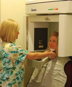
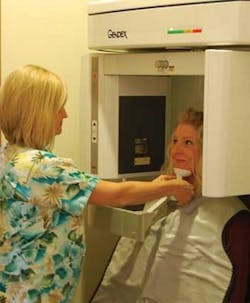
Just when we thought it could not get any better, in-office cone beam imaging became available to boost the convenience of the digital age even more. In-office 3-D scanning technology can increase revenue potential from implants and periodontal therapy without extra hassles for the patient. We can diagnose more predictably from a cone beam scan while avoiding farming out our patients to another venue for the imaging. Patients maintain their comfort zone in our office, and we don't have to wait for radiology reports.
My colleague, Dr. Walter Chitwood, a board-certified implant dentist, has experienced the convenience of cone beam technology firsthand with his GXCB-500™. As for time savings, the scan is completed in 8.9 seconds and reconstructed in less than 20 seconds. In conjunction with his Keystone treatment planning software, he can visualize virtual teeth placement, assess bone density, identify alveolar nerves, and detect implant proximity.
He says, “Because of the efficiency of this imaging method, implant surgery can be scheduled after the first visit, instead of losing time and perhaps case acceptance while patients are shuffled between imaging centers and the office.” He also keeps in touch more easily and efficiently with his referring dentists by printing and mailing his scans or e-mailing them.
Dr. Chitwood and I agree that with all of the complexities of running a growing dental practice, digital imaging maximizes our efficiency, the patient's educational opportunities, and our income — without increasing our workload. When we add convenience to the menu, and take some of the stress off our plates, we get the chance to reap our just desserts — a more successful dental practice.
Dr. Terry L. Myers is a fellow in the Academy of General Dentistry and a member of the Academy of Cosmetic Dentistry and the Dental Sleep Disorder Society. He has a private practice in Belton, Mo. You may contact Dr. Myers by e-mail at [email protected].

