X Files #11 — Yes, we scan — making the best choice in 3–D field of view
by Terry L. Myers, DDS, FAGD
For more on this topic, go to www.dentaleconomics.com and search using the following key words: cone beam, 3–D imaging, fields of view, digital imaging, Dr. Terry L. Myers.
American poet Edwin Markham said, “Choices are the hinges of destiny.” Decisions we make now will shape our practices for years ahead. This month’s column is dedicated to 3–D imaging fields of view. There is no question that 3–D images provide a view that previously could only be achieved under surgical conditions. Cone beam machines are available in small (a few teeth), medium (jaws and condyles), or full (most of the skull) to extended (cephalometric) fields of view. With the increasingly competitive nature of our business, the question is not whether to get a cone beam scanner, but which field of view best suits your practice.
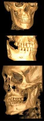
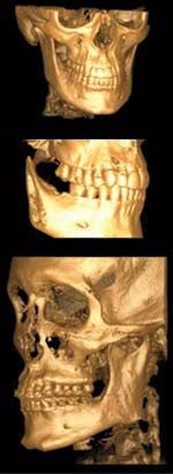
Dr. Bruce A. Howerton, oral and maxillofacial radiologist, 3–D educator, and owner of both medium and full–view systems (i–CAT® and GXCB–500™), explains that certain specialists require the full or extended field of view for diagnosis and treatment planning: “For oral and maxillofacial surgeons, the full field of view offers the information necessary for facial reconstructive and orthognathic surgeries, and orthodontists also need craniofacial anatomical landmarks for treatment planning.”
The scan data from these full and extended views can be brought into other software for accurately predicting results from orthognathic surgery, as well as turned into 3–D computer models for high–tech tooth movement with programs such as SureSmile®.
An endodontist colleague who routinely uses 3–D radiography in private practice, Dr. Ralan Wong, says with the medium field he can compare both sides of the anatomy, something he is unable to do with small field of view without taking more scans. “With medium field of view, I can see the entire arch and symmetry of the dentition. For example, when looking at a first molar, I find that many times if one tooth has four canals, the other side will have four as well.”
Dr. Howerton adds, “The field of view should capture the regions of interest and prevent multiple scans at smaller fields of view.”
As a general dentist, I chose the medium field of view because the scans show just the area where I work. I can still explore the area of the TMJ and into the glenoid fossa. My machine and its sister, the i–CAT, both collimate down to multiple sizes, the lowest being 4 cm in height, so I have choices as to how much anatomy I want to capture. In the few instances where I need to view the whole skull, a software application allows me to take an image of the upper part of the skull and “stitch” the two images together.
As I continue to learn about this topic, my choice makes even better sense. Medium field of view exposes my patients to less radiation. The 8.9–second scan time decreases the incidence of blurred images from patient movement. From the economics side, medium field of view is fantastic, especially for the procedures general dentists and some specialists routinely perform. It made 3–D imaging a possibility for a generalist like me who may not otherwise be able to afford the full–field scanner.
In the past, I had to send my patients to an imaging center or a hospital. Now my patients appreciate being scanned in the comfort of my office. I also gained confidence to expand the number of implant cases and other procedures I wouldn’t try without the 3–D view.
Some dentists are concerned about the possibility of missing a nondental–related pathology on a full–field scan. Since these scans (and in some 2–D X–rays) show more than the teeth and jaws, it’s prudent to understand that dentists can be responsible for information even in areas outside their scope of learning.
I encourage dentists to gain adequate training to properly read 3–D scans, no matter the field of view. Dr. Howerton suggests: “The issue can be solved by having a qualified oral and maxillofacial radiologist review the data for a nominal fee. Very few patients will refuse to pay for such additional information.”
Adding cone beam to my office has improved my practice clinically and financially. Dentists who want to add depth to their practices and take control of their destiny should start considering which field of view best suits their office. Then, when patients and referring dentists inquire about the new technology, they will be proud to answer, “Yes, we scan.”
Dr. Terry L. Myers is a fellow in the Academy of General Dentistry and a member of the Academy of Cosmetic Dentistry and the Dental Sleep Disorder Society. He has a private practice in Belton, Mo. Contact Dr. Myers by e-mal at [email protected].