Maintaining curvatures: strategies and concepts
by Richard Mounce, DDS
For more on this topic, go to www.dentaleconomics.com and search using the following key words: rapidly curving apical anatomy, surgical operating microscope, RNT files, maintaining curvatures.
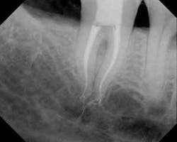
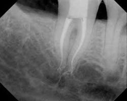
One of the most challenging nonsurgical anatomic entities that a clinician must address is rapidly curving apical anatomy. Canals that are relatively straight in the coronal half of the roots that abruptly curve in the apical half are at risk for ledging, blockage, instrument separation, and transportation of the minor constriction of the apical foramen, among a host of other possible iatrogenic events. Such a tooth is shown in Fig. 1.
Preventing these issues and accenting the creation of ideal canal shapes is addressed here:
1) Access into the pulp chamber and coronal third is the gatekeeper for the apical regions of the tooth. Access is ideally straight line. Access should allow a hand file or rotary nickel titanium instrument to lie in the canal without being deflected off the access walls. Such access provides the clinician with the best possible tactile control.
2) The greater the degree of lighting and magnification the clinician can bring into the apical regions of the canal the better, ideally with the surgical operating microscope (SOM) (Global Surgical, St. Louis, Mo.). Working in a dark hole by tactile feel alone is unnecessary and unproductive.
3) Access debris and pulp tissue are flushed away from the chamber before any instruments are placed into the canals. Clinicians must be sure that all canals are located and the pulp chamber has been fully unroofed before they attempt canal negotiation. The cervical dentinal triangle can be routinely removed using a brush stroke against the root wall of greatest dentin thickness with orifice openers. For vital cases, this use of orifice openers is performed with a viscous EDTA gel in the chamber, such as Slick Gel (SybronEndo, Orange, Calif.) or FileEze (Ultradent, South Jordan, Utah).
4) After these initial steps, the clinician should make sure the canals are patent. I do this by placing a No. 6 hand K file apically to the estimated working length. I note how easily the file is inserted apically and if there is any tactile "pop" at the minor constriction (MC) of the apical foramen. Often, such a pop is present and is a sign that the canal is open, patent, and negotiable.
5) Subsequently coronal and middle third enlargement is carried out. One byproduct of canal enlargement is that irrigants are allowed to flow into the apical third, especially with recapitulation. Apical irrigation and recapitulation are central to the prevention of blockage and maintaining apical control of enlargement. As the clinician moves apically down the root canal system with rotary nickel titanium files, there are three key strategies that allow the apical third of such a curvature to be enlarged safely:
a) The canal should be enlarged to a No. 15 hand K file diameter first. This can be carried out by hand or with an M4 reciprocating handpiece (SybronEndo). RNT files are not placed into this portion of the canal until it has been fully negotiated and such a glide path has been created. When a hand file is removed from a portion of a canal after negotiation and it has a curvature imparted onto it, this tells the clinician what degree of difficulty is present.
b) Irrigation should be copious after every RNT insertion with a side-venting needle. Reciprocation is carried out after every RNT insertion and irrigation to the estimated or true working length. These hand files are precurved before insertion, and the clinician must be sure that the canal is open and passively negotiable before inserting a subsequent RNT instrument. Skipping irrigations and recapitulations is the harbinger of a broken RNT instrument or other iatrogenic event.
c) Coincident to these concepts, the clinician must be aware of the position of the MC of the apical foramen precisely and with each insertion, and not to traverse the MC to avoid iatrogenic outcomes. It is important that the clinician appreciate that the working length will get shorter as the canal preparation proceeds.
It is noteworthy that the treatment shown was performed with .10 and .08 Twisted Files (SybronEndo). Because TF is never ground across its crystalline grain structure due to its fracture resistance and flexibility, it is able to be inserted into apical curvatures in tapers that were not previously possible. TF is used crown down, from larger to smaller tapers with the aforementioned irrigation, recapitulation, and insertion sequence. I welcome your questions and feedback.
Dr. Mounce offers intensive customized endodontic single-day training programs in his office for groups of one to two doctors. For information, contact Dennis at (360) 891-9111 or write [email protected]. Dr. Mounce lectures globally and is widely published. He is in private practice in endodontics in Vancouver, Wash.

