Achieving reproducible, positive clinical outcomes for adjacent Class II composites with a universal adhesive and bulk-fill composite
Above: Dr. Amisha Singh interviews Dr. Nathaniel Lawson about dental materials, his DE article, and more.
NextGen Leadership Showcase
To introduce you to the next generation of dental key opinion leaders (KOLs), DE has launched a special article series. Over the next several months, we will introduce you to emerging KOLs and the innovations they are fostering. You’ll also have a chance to listen to their stories in video interviews. The first article in this series, “Perspectives of dental diversity” by Amisha Singh, DDS, appeared in our January issue. Search “Singh” at dentaleconomics.com to read it. Search “Winter” to read last month’s article, “Smile design with zirconia,” by Sarah Winter, DMD.
Although stunning anterior composites may be the showcase work for a practice, the bread-and-butter procedure of most restorative dental practices is the Class II composite restoration. Proficiency and efficiency in this basic procedure can enhance the productivity of a practice and increase the dentist’s satisfaction with his or her profession. This article will describe the techniques and materials you need to achieve reproducible clinical outcomes for neighboring Class II restorations.
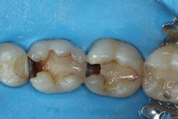
A patient presented with two failing composite restorations on teeth Nos. 30 and 31 (figure 1). A DermaDam rubber dam (Ultradent Products) was placed. This rubber dam was used because it has adequate thickness to displace tissue, yet sufficient compliance to stretch over larger clamped teeth without tearing. Rubber dam placement is used for only about 12% of operative dentistry procedures.1 Placing a rubber dam not only prevents salivary contamination of adhesive bonding procedures, but it also filters discarded dental materials, displaces the tongue and cheek, and reduces dental mirror condensation. The additional time required to place the rubber dam can be easily compensated for by these time-saving advantages, particularly when working on multiple restorations.
Figure 1: Initial presentation of failing composites on teeth Nos. 30 and 31
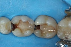
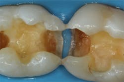
After removing the previous restorations, the distal axial wall of tooth No. 30 and the mesial axial wall of No. 31 had deep caries (figure 2). TheraCal LC (Bisco Dental)—a light-cured, resin-based liner containing calcium silicate (the active ingredient of MTA)—was placed over these areas of deep caries. A thin layer of the material was placed just at the areas of deep caries (figure 3) and light cured. Layers should be kept thin to ensure that the entire bulk of this opaque material cures, and placement should be limited to areas where needed in order to use the surrounding dentin for adhesive bonding. The dentin should be moist but not overly wet when TheraCal LC is applied to ensure that the material “sticks” to the tooth. Because this material is resin based, it is not necessary to cover this material with a separate resin-modified glass ionomer (RMGI) liner.
Figure 2: Deep caries remains on the distal axial wall of tooth No. 30 and the mesial axial wall of No. 31
Ecosite-Bond universal adhesive (DMG America) was used for these restorations. This adhesive can be used in a self-etch or etch-and-rinse mode. A recent systematic review of clinical trials reported that there was no difference in the incidence of postoperative sensitivity between self-etch and etch-and-rinse adhesives in most clinical trials.2 However, the authors of that study noted that these results may not apply to deeper posterior resin composites. In deep dentin, there are more dentinal tubules with a greater diameter. Due to the depth of the Class II preparations in this case, a selective-etch technique was used.
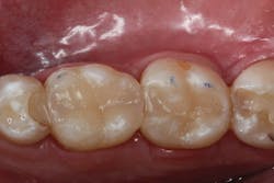
Figure 3: Placement of resin-based, light-cured calcium silicate liner (TheraCal LC; Bisco Dental)
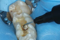
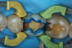
In this technique, phosphoric acid is applied to the enamel margins for 15 seconds (figure 4) and then rinsed off. Phosphoric acid creates an etch pattern on enamel that has been shown to reduce marginal discoloration in clinical trials with universal adhesives.3,4 Then the Ecosite-Bond universal adhesive was applied to the entire preparation, allowing self-etching to occur on dentin. In this way, the Ecosite-Bond universal adhesive achieves the advantages of etch-and-rinse on the enamel and self-etch on the dentin.
Figure 4: Use of a selective-etch technique prior to using the Ecosite-Bond universal adhesive (DMG America)
Prior to filling, the depth of the preparations were measured as greater than 6 mm in the mesial boxes of teeth Nos. 30 and 31. Therefore, at least three increments of conventional composite would need to be placed to ensure that all layers of composite were sufficiently light cured. Each layer of composite that is placed, however, has the potential of trapping voids between layers. If these voids occur at the external margins of the restoration, they can act as plaque traps. Bulk-fill composites are a category of composites that are slightly more translucent, allowing the curing light to cure a 4 mm or 5 mm increment.5
Figure 5: Wide-mouth compule of Ecosite Bulk Fill (DMG America) allows efficient, even extrusion
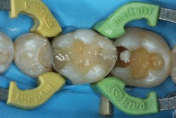
Ecosite Bulk Fill composite (DMG America) was used as it has a wide-mouth compule that allows efficient, even extrusion of material into the preparation (figure 5). This material has a 5 mm depth of cure, but the preparation was deeper than 6 mm, so it was placed in two layers. It is important to first condense the material into the depth of the boxes to make sure there is adequate adaption to the preparation floor. Then, the surface layer of the first increment of composite should be adapted to the walls of the preparation with a round instrument to create a rounded line angle at the interface between the composite and the tooth preparation (figure 6). After a 20-second light cure, the final increment was placed. In order to minimize marginal discrepancies and “white lines,” the composite material should be spread onto the adjacent tooth surface.
Figure 6: Adaptation of the first layer of Ecosite Bulk Fill composite
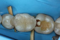
For these neighboring restorations, the choice was made to restore the teeth one at a time using a sectional matrix system (Triodent V3 Ring; Ultradent Products). The advantage of restoring only one tooth at a time is that it is possible to perfect the interproximal contours of the first restoration prior to initiating the neighboring restoration. In this case, the distal marginal ridge of tooth No. 30 encroached into the mesial box of No. 31 after the initial fill (figure 7).
Figure 7: Second layer of composite placed with the marginal ridge of tooth No. 30 encroaching on the box of No. 31
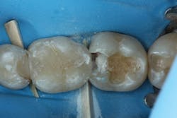
In order to recontour the proximal contact without gauging the composite, a medium grit flexible disc (FlexiDisc; Cosmedent Inc.) was used (figures 8 and 9). Other clinicians prefer to restore both teeth at the same time and attempt to evenly distribute the space by careful placement of the sectional matrices or by holding the place of the unrestored tooth with a ball of Teflon tape.
Figure 8: Proximal contacts adjusted with medium grit flexible polisher (FlexiDisc; Cosmedent Inc.)
After both restorations were filled, the rubber dam was removed and the occlusal contacts were adjusted. The final polish of the restoration was achieved with a Jiffy abrasive polishing brush (Ultradent Products), which is a silicon carbide impregnated polishing brush. The high polish achieved by the Ecosite Bulk Fill composite can be credited to homogenously dispersed nanofillers (figure 10).
Figure 9: Favorable proximal contours achieved on the distal of tooth No. 30
Additionally, the translucency of the composite allows it to blend well with the surrounding tooth structure. The final restorations should have adequate bond and strength to withstand oral function, as well as sufficient polish and contour to give the patient the opportunity to maintain these restorations with proper hygiene.
Figure 10: The final polish and translucency of Ecosite Bulk-Fill composite
References
1. Gilbert GH, Litaker MS, Pihlstrom DJ, Amundson CW, Gordan VV; DPBRN Collaborative Group. Rubber dam use during routine operative dentistry procedures: findings from the dental PBRN. Tex Dent J. 2013;130(4):337-347.
2. Reis A, Dourado Loguercio A, Schroeder M, Luque-Martinez I, Masterson D, Cople Maia L. Does the adhesive strategy influence the post-operative sensitivity in adult patients with posterior resin composite restorations?: A systematic review and meta-analysis. Dent Mater. 2015;31(9):1052-1067. doi:10.1016/j.dental.2015.06.001.
3. Ruschel VC, Shibata S, Stolf SC, et al. Eighteen-month clinical study of universal adhesives in noncarious cervical lesions. Oper Dent. 2018;43(3):241-249. doi:10.2341/16-320-C.
4. Lawson NC, Robles A, Fu CC, Lin CP, Sawlani K, Burgess JO. Two-year clinical trial of a universal adhesive in total-etch and self-etch mode in non-carious cervical lesions. J Dent. 2015;43(10):1229-1234. doi:10.1016/j.jdent.2015.07.009.
5. Van Ende A, De Munck J, Lise DP, Van Meerbeek B. Bulk-fill composites: a review of the current literature. J Adhes Dent. 2017;19(2):95-109. doi:10.3290/j.jad.a38141.
Nathaniel Lawson, DMD, PhD, is director of the Division of Biomaterials at the University of Alabama School of Dentistry.
