Making optimal zirconia crowns
Q: What is the most adequate tooth preparation for a zirconia crown? I see numerous types suggested by companies and speakers. Additionally, I have had some laboratories send zirconia crowns back to me that have been purposely fabricated out of occlusion, which has made the occlusal zirconia very thin. Why are dental technicians not making zirconia restorations in proper occlusion? How thick or thin should the zirconia be on a full-zirconia crown?
A: Some readers will feel that the information I provide is well known by all dentists. That is definitely not the case. An observation of cases sent into dental laboratories shows there are significant problems with crown preparations and impressions!
As you probably know, full-zirconia crowns now dominate the crown market. During the approximately 10 years full-zirconia crowns have been available, their increase in use and clinical success have been phenomenal. The major limitation has been esthetic properties, which have often been less than optimal.
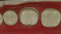
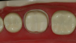
Figure 1: Adequate/optimal prep for full-zirconia or full-metal crowns
Companies producing zirconia have attempted to make its esthetic properties more adequate by adding various percentages of oxides and coloring ions to the powder. The result has improved esthetics somewhat, but it has caused a reduction in strength and transformation toughening. Transformation toughening is a property that makes the ceramic more difficult to crack and break. The overall state of the art on zirconia is gross confusion on the part of dentists and many dental laboratories.
At this time, dentists are well advised to stay with the original tetragonal zirconias, originally introduced in the US for full-zirconia crowns by Glidewell 10 years ago as BruxZir, or other brands using the original tetragonal type of zirconia. When more supportive research becomes available on the many new versions of esthetic or cubic zirconia with higher percentages of oxides, color pigments, and other ingredients, and they are shown to be clinically successful, that will be the time to change to those versions.
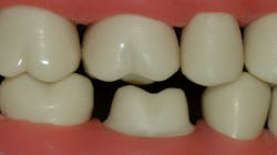
Figure 2: Example of an optimal prep for full-zirconia crowns
A new classification of ceramic restorations related to clinical indications has recently been published and is summarized below.1
• Class 1, porcelains—examples are feldspathic porcelain, low-fusing porcelain, (KIC <1.0 fracture toughness, and 100 or less MPa) inlays, onlays, and veneers adhesively cemented.
• Class 2, leucite glass-ceramics—examples are Vita Mark II (Straumann), IPS Empress (Ivoclar Vivadent, KIC >1.0 fracture toughness, and >100 MPa) single-unit anterior or posterior adhesively cemented.
• Class 3, lithium disilicate—example is IPS e.max
(Ivoclar Vivadent, KIC >2.0 fracture toughness, and >300 MPa) single-unit or three-unit anterior.
• Class 4, cubic-containing zirconia—examples are cubeX2 cubic zirconia (Dental Direkt), Katana STML/UTML (Kuraray Noritake), Lava Esthetic (3M Oral Care, KIC >3.5 fracture toughness, and >500 MPa) three-unit anterior or posterior.
• Class 5, tetragonal zirconia—examples are original BruxZir, Lava Plus (3M, KIC >5.0 fracture toughness, and >800 MPa) four or more units anterior or posterior.
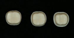
Figure 3: An adequate prep for full-zirconia or full-metal crowns, which are the same.
A word to the wise
Consult with your technician. Make sure your lab is using an FDA-cleared zirconia. Such zirconias must pass ISO and American Dental Association standards as stated in the above classification. Technicians and dentists are encouraged to become conversant and knowledgeable about the five categories of ceramics to facilitate their informed clinical choices.
Challenges observed in labs relative to zirconia restorations
Inadequate tooth preparations. In answer to your question, the preparation recommended is for the original tetragonal zirconia. Long-term clinical research in the Technologies in Restoratives and Caries Research (TRAC) division of the nonprofit Clinicians Report Foundation has found no breakage of single tetragonal zirconia crowns in the nine-year study.2 Research indicates that most or all current zirconia formulations should be able to serve adequately using the following tooth preparation characteristics.
Look at the photos of adequate tooth preparations in Figures 1–5 as you read the prep characteristics.
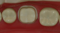
Figure 4: Porcelain-fused-to-metal (PFM) crown prep
Many of the zirconia companies describe minimal depth preparations—not optimal preparations. As a result, many zirconia crown preps observed in labs are too shallow. Figures 1–3 show an adequate/optimal prep for full-zirconia or full-metal crowns, which are the same.
Note these characteristics for adequate/optimal preps for zirconia crowns:
• The gingival margins should be at least 0.6 mm deep.
• All of the axial walls should be at least 1.0 mm deep.
• The occlusal cuts should be anatomic following the original tooth occlusal anatomy and at least 1.5 mm deep.
Why should zirconia preps be this deep?
Many labs are relieving the occlusal zirconia by 0.3–0.5 mm (300–500 microns) to ensure that you will not complain about the crowns being too high and to minimize chairside cutting of the ceramic that can risk crown fracture. I strongly disagree with this technique, since the out-of-occlusion tooth requires months to extrude, and the forces on adjacent teeth often break the cusps of those teeth. In my opinion, the spacing should be about one-tenth of what we are seeing coming from many labs or about 30–50 microns. That is roughly the thickness of a human hair, and natural tooth extrusion will usually bring the crown into occlusal contact in a few weeks.
Assuming the lab technician relieves the occlusal 0.5 mm, you now have only 1 mm of zirconia remaining on the occlusal. Now, put minimal anatomy into the occlusal surface of the crown, and your occlusal crown thickness will be about 0.6–0.5 mm, which is considered to be the minimal amount of material for strength.
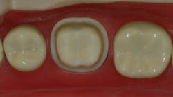
Figure 5: PFM crown prep axial walls should be slightly deeper than for zirconia or metal
The remaining Figures 4 and 5 are shown for comparison with the zirconia photos. As is evident from the photos, the porcelain-fused-to-metal (PFM) crown prep axial walls should be slightly deeper than for zirconia or metal (1.5 mm) to accommodate 0.3–0.5 mm of metal substructure and the fused or pressed ceramic veneering material. The occlusal reduction should be at least 1.5 mm and preferably 2.0 mm for the same reason.
An adequate prep for IPS e.max is even deeper (figure 5). It is evident that the cervical cuts should be at least 1.0 mm deep, and the occlusal reduction is best at 2.0 mm to allow adequate thickness for full strength of this glass-ceramic and occlusal anatomy.
Inadequate impressions. A high percentage of impressions, both conventional and scanned, do not show margins adequately. Large labs estimate 70%–90% of impressions could be better. Some of the techniques promoted for soft-tissue management are unpredictable, and technicians must fake at least some portion of the margins on most conventional or digital impressions. When doing this, the margins do not fit. Margins must be visible to the naked eye, or they cannot be scanned or recorded on vinyl or polyether. I strongly recommend that subgingival margins require either a standard two-cord technique or a modified one-cord technique.
The proven, most-predictable impression procedure for subgingival crown margins:
• Prep to the gingival line, with no blood stimulated. Avoid the gingiva.
• Place a first cord to fill half of the sulcus.
• Prep to the coronal limit of the first cord.
• Place a second cord with styptic on it and wait a few minutes.
• Remove the second cord.
• Leave the first cord in place.
• Make the impression.
• Remove the first cord.
An alternate proven technique for impressions of subgingival margins:
• Prep to the gingival line, with no blood stimulated. Avoid the gingiva.
• Place a first cord to fill half of the sulcus.
• Prep to the coronal limit of the first cord.
• Place a styptic-impregnated paste (examples are 3M Oral Care Astringent Retraction Paste, Parkell Dryz Gingival Retraction Paste, Acteon Group Expasyl Gingival Retraction Material, Premier Dental Traxodent Hemodent Retraction Paste) and wait a few minutes.
• Wash out the styptic paste.
• Leave the first cord in place.
• Make the impression.
• Remove the first cord.
There are undoubtedly other successful alternative tissue-management techniques. Look at your impressions. If you can see every aspect of the gingival margins clearly—without compromise—stay with your current procedure. If you have to fake some margins, change your technique! The well-known fact is, currently, that most dentists are not making adequate impressions.
Summary
The zirconia revolution has brought conceptual and technique challenges, but, so far, the data show zirconia is working well clinically. We dentists are the challenge. Look at your tooth preparations. Do they have the characteristics shown in this article? Evaluate your impression procedure. Is it acceptable every time? Learn the characteristics of the ceramics described, and both you and your patients will have successful restorations.
References
1. Morris G. Use ADA-approved ISO standards to confidently recommend all-ceramic esthetic materials. J Dent Technology. 2018;6:22-24.
2. Zirconia: most durable tooth-colored crown material in practice-based clinical study. Clinicians Report. 2018;11(11):1-3.
Author’s note: The following educational materials from Practical Clinical Courses will offer you and your staff more insight on the topic discussed in this question.
One-hour videos:
- Crowns—Materials and Techniques for the Best Results (Item No. V1987)
- Foolproof, Fast Single-Crown Procedure (Item No. V1980)
Two-day hands-on courses:
- Restorative Dentistry 2—Fixed Prosthodontics with Dr. Gordon Christensen
- Implementing Cone-Beam Imaging into Your Dental Practice with Dr. Dale Miles and Dr. Gordon Christensen
For more information about these educational products, call (800) 223-6569 or visit pccdental.com.
Gordon J. Christensen, DDS, PhD, MSD, is a practicing prosthodontist in Provo, Utah. He is the founder and CEO of Practical Clinical Courses, an international continuing education organization founded in 1981 for dental professionals. Dr. Christensen is cofounder (with his wife, Rella Christensen, PhD, RDH) and CEO of Clinicians Report.