Sticking to the rules: Clinical success with universal adhesives
Ron Kaminer, DDS
When one mentionsadhesives in dentistry, the first thing that comes to mind is the term generation. Adhesive dentistry has changed dramatically since the early work of Michael Buonocore, DMD, MS.1 As the dentistry has changed or improved, so has the chemistry, the delivery, the mechanics of the technique, as well as the effectiveness of the product. Today, most dentists use the latest generation of adhesives, classified as universal adhesives.
Universal adhesives allow dentists to use the product in a variety of modes—i.e., self-etch, selective-etch, and total-etch—with no difference in outcome. While the pH of these adhesives tends to be on the lower side (2.2–3.4), which is more than sufficient for dentinal bonding, the concern for many lies in the ability to effectively etch and bond to enamel with these products.2 While many continue to use these products in a self-etch mode and have advocated selective-etch for years, this remains the technique of choice.
The selective-etch technique involves etching the enamel with phosphoric acid prior to using the universal adhesive. Newer studies recommend shorter etch times, with some studies touting no benefit after a three-second phosphoric acid etch.3 Most also recommend some alteration to the enamel, as in a bevel, since universal adhesives bond better to cut enamel versus uncut enamel.4 Adhesives should be massaged thoroughly into the tooth surface for optimal penetration into the dentinal tubules.5
Universal adhesives pack a tremendous amount of chemistry into one bottle. The fragility of the monomers comes into question for many in these one-bottle systems, although the stability of the chemistry has improved dramatically throughout the years. When using a one-bottle system, one should be cautious to recap the bottle immediately after use to minimize any potential evaporation of the material from inside the bottle.
One interesting product that I have used for a long time is Futurabond U (Voco), which has a blister pack delivery system to reduce material evaporation. Another feature to note about Futurabond U is the ability to use this material in an indirect fashion. Some universal adhesives need an additional component (dual-cure activator) when used with an indirect restoration. Futurabond U has dual-cure capabilities already built in, which eliminates a step when bonding in a post, cementing a crown, or cementing an onlay.
Case No. 1
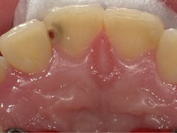
This case involved a seemingly routine Class III cavity on a 13-year-old girl, who was terribly apprehensive of the dentist and injections (figure 1). After initial consultation with the patient and her parent, it was decided that we would use the LiteTouch Er:YAG laser (AMD Lasers) instead of a high-speed handpiece for the cavity preparation. This laser can often be used without anesthetic, as its energy is absorbed by water and hydroxyapatite to cut hard tissue in an efficient manner. The laser’s energy will also remove the smear layer, making it ideal for a bonded restoration.6
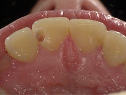
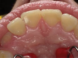
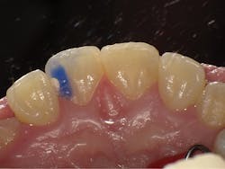
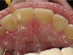
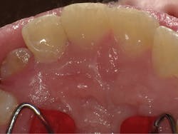
The completed preparation shows clean margins and complete decay removal (figure 2). Due to the depth of the preparation, a urethane dimethacrylate calcium liner (Calcimol, Voco) was placed and light-cured for 15 seconds (figure 3). The enamel margins were etched for 3 seconds (Vococid, Voco) and the etch was rinsed off (figure 4). A mylar strip and wedge (Composi-Tight 3D Fusion Wedge, Garrison Dental Solutions) were placed and Futurabond U was applied for 20 seconds and massaged throughout the preparation, air-dried, and light-cured (figure 5). Admira Fusion (Voco) shade A2 was placed in two increments and light-cured. The final polished restoration shows ideal esthetics (figure 6).
Figure 1: Preop: Class III cavity on 13-year-old patient
Figure 2: Completed preparation with clean margins and complete decay removal
Figure 3: Due to the depth of the preparation, Calcimol was placed and light-cured for 15 seconds.
Figure 4: Enamel margins were etched with Vococid for 3 seconds, and then etch was rinsed off.
Figure 5: Composi-Tight 3D Fusion Wedge was placed. Futurabond U was applied for 20 seconds and massaged throughout the prep, air-dried, and light-cured.
Figure 6: The final polished restoration shows ideal esthetics after Admira Fusion shade A2 was placed in two increments and light-cured.
Case No. 2
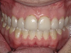
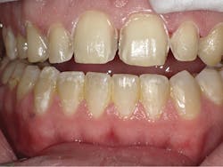
This patient presented for a smile makeover due to failing old veneers. The preop photo shows bulky veneers with bacterial stain at the margins and poor esthetics (figure 7). The veneers were removed using an electric high-speed handpiece (NSK) and a variety of diamond burs (Microcopy USA; figure 8). The preparations were smoothed off, impressioned, and an esthetic provisional was fabricated.
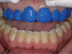
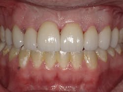
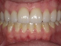
On the insertion appointment, the provisional was removed and the preparations were cleaned with a chlorhexidine cavity cleanser (Cavity Cleanser, Bisco). The veneers were silanated, and the preparations were etched with 37% phosphoric acid for 10 seconds (Vococid; figure 9). The etch was rinsed off, the preparations dried, and Futurabond U was applied to all the preparations following the manufacturer’s instructions. The universal adhesive was cured for 20 seconds on each preparation. A clear sheen after curing indicates complete coverage of the preparation with the adhesive (figure 10). The veneers were bonded into place using a light-cured resin cement (Choice 2, Bisco). The margins were finished with a combination of finishing carbides and diamonds (Microcopy USA; figure 11). A nine-month postoperative photo depicts ideal gingival health and outstanding esthetics (figure 12).
Figure 7: Preop: Smile makeover case due to failing veneers. Note bulky veneers with bacterial stain at margins and poor esthetics.
Figure 8: Veneers were removed with NSK electric high-speed handpiece and Microcopy diamond burs.
Figure 9: Veneers were silanated, and preps were etched with 37% Vococid for 10 seconds.
Figure 10: After etch was rinsed off and preps were dried, Futurabond U was applied to all preparations. Each prep was cured for 20 seconds. A clear sheen after curing indicates complete coverage.
Figure 11: Margins were finished with a combination of Microcopy finishing carbides and diamonds.
Figure 12: Nine months post-op: Ideal gingival health and esthetics
With more than 1,000 direct and indirect restorations spanning many years, I have seen universal adhesives stand the test of time. Long-term success has been achieved in routine restorative dentistry as well as complex crown-and-bridge dentistry. With careful adherence to manufacturers’ instructions, you can achieve the same success as well and be confident in your adhesive dentistry procedures.
References
1. Buonocore MG. A simple method of increasing the adhesion of acrylic filling to enamel surfaces. J Dent Res. 1955;34(6):849-853. doi: 10.1177/00220345550340060801.
2. Rosa WL, Piva E, Silva AF. Bond strength of universal adhesives: A systematic review and meta-analysis. J Dent. 2015;43(7):765-776. doi: 10.1016/j.jdent.2015.04.003.
3. Tsujimoto A, Barkmeier WW, Takamizawa T, Latta MA, Miyazaki M. The effect of phosphoric acid pre-etching times on bonding performance and surface free energy with single-step self-etch adhesives. J Oper Dent. 2016;41(4):441-449. doi: 10.2341/15-221-L.
4. Swanson TK, Feigal RJ, Tantbirojn D, Hodges JS. Effect of adhesive systems and bevel on enamel margin integrity in primary and permanent teeth. Pediatr Dent. 2008;30(2):134-140.
5. Loguercio A, Muñoz MA, Luque-Martinez I, Hass A, Reis A, Perdigão J. Does active application of universal adhesives to enamel in self-etch mode improve their performance? J Dent. 2015;43(9):1060-1070. doi: 10.1016/j.jdent.2015.04.005.
6. Kataumi M, Nakajima M, Yamada T, Tagami J. Tensile bond strength and SEM evaluation of Er:YAG laser irradiated dentin using dentin adhesive. Dent Mater J. 1998;17(2):125-138. doi: 10.4012/dmj.17.125.
Ron Kaminer, DDS, a 1990 graduate of the State University of New York at Buffalo, is a prolific author, lecturer, and expert on dental lasers. He is a clinical consultant/lecturer for numerous companies (GC America, AMD Lasers, SDI, Camsight, NuCalm, Solutionreach), maintains two teaching appointments, and serves on the editorial board of Dental Product Shopper. He maintains practices in Hewlett and Oceanside, New York.
