Overcoming CMOS sensor challenges
Each month, Dr. Gordon Christensen answers a question from our readers about everyday dentistry.
Q: I have been practicing for many years and have lived through the analog radiograph era as well as the introduction, use, and current state of dental digital radiographs. I like certain aspects of the currently available complementary metal oxide semiconductor (CMOS) sensor concept, but the limitations far outweigh the advantages. Can’t we do better than this? A day does not pass when I don’t complain about some aspect of the current CMOS digital sensors. What is in the future for this major void in dental practice?
A: Many older dentists have had experience with analog radiographs, which are not used to a significant degree at this time. They had numerous disadvantages, including the need to maintain a darkroom and developing area, deal with developing and fixing solutions, purchase and change the solutions routinely, organize and save the images, and tolerate patients’ worry about the relatively high radiation required with their use.
But the analog radiographs were thin, flexible, easy to place and use on a positioning device, easy to place in the mouth without causing patient discomfort, relatively inexpensive, and they actually showed initial caries relatively well.
What are the complaints dentists make concerning the current digital CMOS sensors and the images they make? The sensors or the images are:
- Excessively expensive
- Thick and resist proper placement
- Rigid and unable to bend to accommodate oral anatomy
- Often painful to use because of their size, rigidity, and sharp angles
- Delicate and break often, especially cords
- Expensive to repair
- Contaminated when used and require routine infection control procedures
- Sometimes difficult to produce predictable, consistent images
- Prone to digital manipulation, perhaps allowing dishonesty
- The same in each brand with changes for different outside coverings, variation in thicknesses, and shapes and angles on the edges
- Basically the same regardless of brand name since all brands use the CMOS sensor concept
- Not equal to the size of the sensors, so a significant portion of the anatomy is not shown on the edges of the sensor
And the big ones:
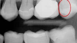
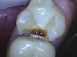
- Aren’t capable of showing initial or even deep dental caries (figures 1 and 2)
- Aren’t expected to be improved or replaced in the near future
Are there advantages to digital CMOS radiography?
Yes, there are a few:
- Fast to use
- Do not require a darkroom and conventional developing
- Have low radiation compared to analog radiographs
- Do not require developing or fixing solutions
- Allow immediate viewing
- Images can be stored and retrieved at will
- Allow easy patient education
What are the alternatives to CMOS digital sensors?
The following concepts are proven adjunctive techniques for detecting dental caries:
Air polisher (Prophy Jet, KaVo, or similar)—to clean the occlusal grooves of stain and plaque. If color remains in the groove, the likelihood of class I caries is very high.
Logicon (if you have Carestream or Kodak sensors only) to determine presence and activity of class II and III areas.
Microlux Transilluminator to determine the presence of class II and III caries.
Silver diamine fluoride to stain caries black in occlusal grooves, easily showing class I caries.
CariVu to show the location and depth of class II and III caries.
Digital Doc LUM to show the location and depth of class II and III caries.
CamX Spectra Caries Detection Aid to locate class I caries.
SoproLife and SoproCare to identify class I caries.
Canary System to locate class II caries.
There is another very good alternative—the use of phosphor plate digital radiographs.
As you know, CMOS and CCD (charge-coupled device) digital sensors have been used in dentistry for many years, and currently CMOS sensors are the most popular. But the use of phosphor plate digital sensors is a viable alternative to replace CMOS sensors for some clinical situations. This technology has been available for many years.
The most popular brands are ScanX from Air Techniques and Soredex Digora Optime from KaVo. Over the past several years, phosphor plate sensors have nearly replaced the previously dominant CMOS sensors in numerous European and Asian countries.
These are the advantages of phosphor plates:
- After purchase of the processor/reader to convert the latent image on the plate to digital, the cost of the sensors is very low and can be used up to 200 times.
- Thinness
- Flexibility similar to film
- Low radiation needed
- Images are equal to but not better than CMOS images
- The image captured is the same size as the sensor, which is much larger than typical CMOS sensor images.
- Simplicity and ease of use
- Sensor positioning for the image is fast and easy, much like film.
- There are five sizes of sensors.
There are several limitations of phosphor plates:
An immediate image is not obtained. A short time is required to convert the latent image on the plate to digital in a processor. With experience, this time is short—just a minute or two. However, this limitation is a challenge when an immediate image is desirable during complex endodontic treatment, impactions, and implants.
While sensors can be used about 200 times before replacement and cost is less than $100 USD, they must be replaced when they develop artifacts, including lines, across the sensor. It is a limitation to have to replace sensors frequently.
The sensors are light-sensitive and must be protected from light exposure.
Using both CMOS sensors and phosphor plates is a logical concept
Some dentists are using phosphor plates for situations not requiring an immediate image, such as dental hygiene appointments and initial exams, and they’re using CMOS sensors for difficult endodontic procedures, removal of impactions, and implant placement. The same dental x-ray unit can be used for both concepts.
How to overcome radiographic challenges in a typical general practice
Radiographs are taken on almost every patient during almost every appointment. They are essential for many oral diagnostic and treatment procedures. Various surveys report diverse estimates of the number of radiographs made in dental practices. It is obvious that hundreds of images are made monthly in a typical busy practice.
I consider the inadequacies of the current generation of digital sensors to be among the worst voids in everyday oral care. What are the major clinical radiographic challenges?
Lack of ability for current digital sensors to show initial dental caries. Clinicians have to wait too long before restoring teeth, which weakens the teeth, requires more time for the procedure, uses more material, and results in more difficult restorative procedures. Apparently, this problem cannot be overcome with the current generation of digital radiographs. Use of caries-detecting devices and careful magnified visual observation are the only current solutions.
Inconsistency of diagnostic data on images. Making images of a specific anatomic area often provides information that leads to different diagnoses of the same situation. Dentists admit that a definitive diagnosis often cannot be made because the radiographs differ in the digital images shown. Making more than one image of a specific suspicious location is one useful solution, but careful clinical visual observation with magnification is usually necessary.
Difficulty properly positioning the sensors. Positioning in both a sensor-holding device and in the mouth are nearly impossible procedures to accomplish due to the thick, rigid, and often painful CMOS sensors. These challenges can be overcome by adding phosphor plate sensors to your practice. Phosphor plate sensors are as easy to use as film, and you can still use CMOS sensors for situations that require an immediate image.
Summary
The transition from analog radiography to digital radiography has brought about some advantages, but most dentists agree that these advantages are outweighed significantly by the limitations of this concept. Until improvements in digital dental radiography occur, this article outlines several ways to mitigate the challenges presented by the current generation of digital radiographs.
Author’s note: The following educational materials are available from Practical Clinical Courses to help you with oral radiography.
One-hour videos:
- Optimizing Digital Radiography (Item V1119)
- Implementing Cone Beam CT Imaging into Your Practice (Item V1174)
Two-day hands-on courses:
- Restorative Dentistry 2—Fixed Prosthodontics with Dr. Gordon Christensen
- Restorative/Implant Dentistry 3 with Dr. Gordon Christensen
For more information about these educational products, call (800) 223-6569 or visit pccdental.com.
GORDON J. CHRISTENSEN, DDS, PhD, MSD, is a practicing prosthodontist in Provo, Utah. He is the founder and CEO of Practical Clinical Courses, an international continuing education organization founded in 1981 for dental professionals. Dr. Christensen is cofounder (with his wife, Rella Christensen, PhD, RDH) and CEO of Clinicians Report
About the Author

Gordon J. Christensen, DDS, PhD, MSD
Gordon J. Christensen, DDS, PhD, MSD, is founder and CEO of Practical Clinical Courses and cofounder of Clinicians Report. His wife, Rella Christensen, PhD, is the cofounder. PCC is an international dental continuing education organization founded in 1981. Dr. Christensen is a practicing prosthodontist in Provo, Utah.