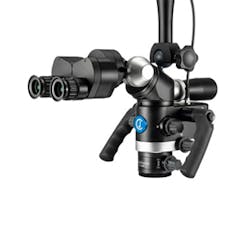
Ergonomics and the dental microscope workflow: How you use it matters

In my years teaching and from the images and videos shared on social networks, I’ve concluded that the dental microscope is being used incorrectly by many dentists. The microscope calls for the operator to work in the neutral seated work posture.1 It allows the operator to see better through multiple magnification steps while recording videos in real time. Yet it is used only for a few specific moments of dental treatment—for the endodontic opening of a root canal, to search for the MB2 canal, to detect a fracture line, to inspect a margin after dental carving to prepare crowns, or to see if a soft tissue suture is properly placed.
All microscope benefits exponentially improve health, control, precision, and communication by the operator and patient health outcomes. The benefits will be higher if it’s used systematically most of the working time.2 The main factors that hinder the proper use of the microscope are work habits/methodology and microscope settings, which interfere with fluidity and comfort during treatment. I’ll discuss these problems to determine why and to try to find simple solutions for its proper use.
Dentist work habits and methodology
Regardless of the type of microscope and accessories, if work habits aren’t adequate, the use of the microscope will be affected since many movements, postures, and forms of work interfere with its proper use. Related factors are position, equipment and furniture, four-handed dentistry, type of microscope stand, operator work position, patient position, and use of a mirror.
Posture
In addition to being a precision tool, the microscope is a tool that, if used properly, provides health to the operator by regulating the working distances and keeping the operator's posture upright to avoid the forward posture that causes musculoskeletal disorders.3,4 Proper use should be guided by knowledge and awareness of the biomechanics of the body (Table 1).

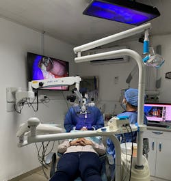
The microscope should be used according to the operator's neutral working posture. The head should be in line with the shoulder and the shoulder with the hips, the hips should be higher than the position of the knees. The forearms should be parallel to the floor or 10 to 15 degrees upward. This position will be determined by the patient's mouth lying on the chair.
The patient's position influences the position of the microscope and the operator head. It is recommended that the binoculars be angled more than 90 degrees slightly to generate a position of the patient's head that promotes operator neutral ear-aligned-shoulder position.5
Equipment and mobiliary
There is furniture with ergonomic characteristics, such as movable carts and delivery systems, that aim to optimize four-handed work and reduce horizontal reach distances. The ergonomic operator's chair promotes the position of the hips higher than the knees, offers convex support in the lower back, and armrests for support in the extremities.6
Four-handed dentistry
The microscope involves the assistant for four-handed dentistry since the operator must focus on the visual field of work through binoculars or screens (3D scope). This leaves the work of distribution and disposition of the external instruments to the assistant. This also generates an economy of unnecessary movements on the part of the operator and a greater organization on the part of the assistant.7 Training the assistant for fluid and efficient work will allow the operator to focus on the visual field of work with the least physical and mental fatigue.
Selecting the microscope stand
The microscope is an ergonomic tool. Incorporating it into the office generates an immediate change in the work design compared to naked-eye dentistry. The team incorporates the microscope according to the dimensions of the office and chooses whether to place it in a wall, ceiling, or stand-alone microscope. Improper selection can obstruct the assistant from doing their job efficiently. For example, the microscope stand has a wide base that can block access to surrounding work areas in a small office. Before acquiring a microscope, consider the dimensions of the office to determine the type of stand, as well as the design of the furniture that facilitates four-handed work.
Dental operator work position
The dental microscope stimulates dental practice at 12-11 o'clock,8 which offers better body symmetry and less compression of joint structures. It also generates a positional change in the operator, who usually tends to turn their head and tilt it to the right and down in a prolonged and repetitive manner throughout the procedure.
Take the time to position the patient based on the neutral posture of the operator. If the patient is not lifted high enough the microscope will be placed too low, affecting the position of the operator's head and the microscope binoculars. If the chair is too high, the microscope will be too high, and the operator's head will be in prolonged hyperextension. If the back of the chair is tilted, this will affect the position of the microscope in the same way, affecting the posture of the lumbar, cervical, and wrists in prolonged extension (figures 1A and 1B).

Use of the mirror
Since most of the work under the microscope is done with indirect vision, the skillful use of the mirror is of great support. With translation or rotation movements of the mirror you can work all the quadrants and tooth surfaces without disturbing the neutral posture of the operator.9
Use dental microscope properly, get all the benefits
A common excuse among those who don’t use microscopes is that the learning curve is long, and they waste time on procedures they can do more quickly without it. If the objective is to be sure that everything is excellent, any doubts of working with the naked eye or limited magnification become certainties when the same work is performed under a microscope, which provides adequate light and maximum magnification. The details "lost" in one are details "gained" visually in another.10
The learning curve and speed of treatment will be achieved with daily use and adequate training in the microscope. It will soon feel automatic to the operator. It’s like any new technology or activity, and that effort is ultimately worth it when it becomes unconscious and the desired speed is gained.
Dental magnification
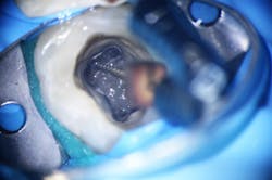
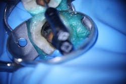
The abuse of high magnification generates a limited visual field that causes a loss of crucial information. Axial axis of the piece and soft tissue information are lost. Try to work with low or intermediate magnifications and leave high magnifications for inspection and diagnosis. The higher the magnification, the smaller the visual field and the higher the resolution. The lower the magnification, the greater the visual field and the lower the resolution (figures 2 and 3).
Attachments that provide ergonomics
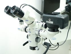
The two most important attachments are the binocular extender (figure 4), which encourages the operator to maintain posture and not look straight ahead for the binoculars, and the multifocal or variofocus lens (figure 5), with a working distance of 200 to 400 mm. This accessory most flattens the learning curve by making the search for working distances less critical than the lens with a fixed focal length.
If we have a tool that gives us so many benefits, these benefits affect a healthy, efficient, and profitable career in the long term. This should be an incentive for proper use and training to alleviate the learning curve. Our bodies and patients will appreciate it.
References
1. Bud et al. The advantages of the dental operative microscope in restorative dentistry. Med Pharm Rep. 2021;94(1):22–27.
2. Low et al. Magnification in endodontics: A review of its application and acceptance among dental practitioners. Eur J Dent. 2018;12(4):610-616
3. Leggat PA, Smith DR. Musculoskeletal disorders self-reported by dentists in Queensland, Australia. Aust Dent J. 2006;51:324–327.
4. Hamann C, Werner RA, Franzblau A, Rodgers PA, Siew C, Gruninger S. Prevalence of carpal tunnel syndrome and median mononeuropathy among dentists. J Am Dent Assoc. 2001;132:163–170.
5. Valachi. Practice Dentistry Pain-Free. Evidence Based Strategies to prevent pain and extend your career. Posturedontic Press; 2008
6. Garcia-Vidal. The Combination of Different Ergonomic Supports during Dental Procedures Reduces the Muscle Activity of the Neck and Shoulder. J Clin Med. 2019;8(8):1230.
7. Finkneiner. Four-Handed Dentistry Revisited. J Contemp Dent Prac. Vol. 1, No 4, 2000.
8. Blanc et al. Variability of Musculoskeletal Strain on Dentists: An Electromyographic and Goniometric Study. Int J Occupational Safe Ergon. 2014;20(2):295–307;144–153.
9. Jin Jeong et al. The effect of indirect vision skills on head and shoulder posture amongst Korean dental hygienists. Eur J Dent Educ. 2020;24(1):17-25
10. Carlos Murgel. Microdentistry, concept, methods and clinical incorporation. Int J Microdent. 2010;2:56-63. https://vittrea.com/producto/microscopio-flexion-basic-de-cj-optik/
11. Carr et al. The use of the operating microscope in endodontics. Dent Clin North Am. 2010;54(2):191-214.
About the Author

Juan Carlos Ortiz Hugues, DDS, CEAS
Juan Carlos Ortiz Hugues, DDS, CEAS, is a master of the Academy of Microscope Enhanced Dentistry (AMED), associate professional in ergonomics by BCPE, CEAS II, and president of the AMED. He wrote the book, Ergonomics Applied to Dental Practice, available from Quintessence Publishing. He provides lectures, training, and advice on advanced dental ergonomics in Latin America and the United States, mainly related to ergonomics applied to dental microscopy.