Digital dental photography - for dentists
The world of dental digital photography, or DDP, is one of those avenues many unsuspecting C.E. travelers have taken only to find that their new cameras, ideas, and techniques are too complex to be implemented into their practices. Most of us are dentists who want to do digital photography - not photographers who make a living doing dentistry. This is not to say there isn’t money in doing digital dental photography. We’ve all either seen or heard about patients turning needed dentistry into wanted dentistry after viewing their dental conditions in sharp detail. Most practices that have adopted digital photography would never go back to film and wait for images to come back from a processing lab.
Psychologically, DDP becomes much more usable as one removes the stigma associated with general film photography and its complexities. There are only a few settings to consider on today’s high-end cameras in order to acquire superb images. In addition, very little knowledge of computers or cameras is required to properly manage the images.
My referring periodontist asked me to help him with his DDP. He had taken a well-known course from a reputable dentist on the lecture circuit, but like many of those who attended, he returned to his office mired in a backlog of treatment and neglected to apply his new knowledge. Like dental treatment, DDP requires repetition for it to be useful in the long term. Even though it requires some training, everyday use during patient treatment will galvanize DDP into your armamentarium.
Choices and applications
The typical high-end single lens reflex, or SLR, digital camera comes in two basic formats. The most misunderstood division of these two types is in their applications, not their designs. The first type is the histogram-based camera. It requires the operator to have a more in-depth understanding of photography and image manipulation because the images are viewed in a graph form that can be altered via software programs such as PhotoShop. Image manipulation is done with this software, which also can be applied to general photography. It relies more heavily on the operator selecting the correct settings and using them properly to capture the ideal image.
The second type of digital camera widely used in dentistry is the through-the-lens, or TTL, metering-based system. The TTL camera relies more upon itself and less upon the operator to acquire images. The advantage of the higher-end TTL cameras is less need to adjust the images because they are automatically adjusted by what the camera “sees.” The question of the camera delivering a “true” image is somewhat controversial, for example, whether the color-and-white balance is an exact replica of the image desired.
I have been using the TTL system for more than four years and have put my total trust in its ability to meter images by itself. Using this camera for shade matching can greatly simplify an otherwise difficult, if not impossible, procedure. Perhaps with the histogram-based system, one may be able to more closely match hue and chroma, but value seems to be the most important aspect for matching shades. The high-end TTL cameras work superbly for this, even though they are based on an automatic metering system. Using a TTL also simplifies teaching digital photography to others. Its implications are emphasized as opposed to histogram manipulations. In addition, the time given to image acquisition is more important to most dentists as compared to image manipulation, which is done after the patient leaves the office.
After attending other DDP lectures, their “wow” factors faded when attendees found out how long it took to re-create that type of dental photography. Many if not most of the genuine experts in DDP spend hours in front of their computers creating beautiful images. As in every facet of dentistry, the proficiency desired depends on how much time one wants to spend in perfecting that specific facet. Granted, DDP can be a great sales tool for case presentation and acceptance, but the additional time spent acquiring the perfect image that needs no software to correct inadequacies will be well worth it. Moreover, using DDP on nearly every patient and procedure will accelerate your proficiency in the acquisition phase. You will then need less proficiency - and time spent - in the manipulation phase after the patient leaves. There is rarely a need to alter images at that point.
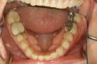
I have read many articles in dental periodicals that often are accompanied by poor-quality images. An obvious example of this is in the use of full-arch mirror images without using lip retractors. It isn’t always easy to acquire images during dental procedures, but it only requires a few more seconds to move the patient’s lips out of the picture (see figures 1 and 2). These images illustrate proper retraction while using an occlusal mouth mirror for full-arch shots.
Another frequent observation is that images are reversed either horizontally or vertically with respect to the desired orientation. This may be caused by an error on submission of the original photos or an error by the publisher. Patients see things so much differently than we do as dentists. When presenting treatment to the patient, it’s more advantageous to orient the images as if the patient is looking into a mirror (see figures 3 and 4). This manipulation is simple with software such as ThumbsPlus and Image FX. Just as in fine restorative dentistry, paying closer attention to details can prevent these simple mistakes.
You're not taking "snapshots"
One of the most absurd notions is that quality images can be acquired with simple point-and-shoot-type cameras. It’s virtually impossible to create decent dental photography, particularly intraoral images, without a ring flash, dual flash, or at the very minimum, a single-point flash system. Some extraoral images can be acquired with point-and-shoot cameras, but some of the photographs found in dental journals with severe and distracting shadows indicate the image was taken without any regard to proper lighting. Higher-end camera and lighting systems often dictate the level of image quality possible, however, several systems produce excellent images for much lower cost than one would expect. Also, as dentists upgrade their equipment, used systems may be found that work beautifully for a lot less money than new ones.
So, what type of flash system is best? Years ago, I purchased a single-point flash that was “state-of-the-art” for intraoral photography, or so I thought. Later, many dentists went to a ring flash because it flooded the area with enough light to acquire high-quality images even far back in the mouth. Consider however, that light correlates to what we are able to see but not necessarily the result we may need. An example of this is the dual-point flash. If we are looking at the subject with both eyes, i.e. using stereoscopic vision, a dual-point flash works well for the anterior segment of the mouth. So it makes sense to use a dual-point flash when acquiring an image for shade matching.
Over time, we found that dental photography is more than just taking pictures of teeth. Another, more general type of illumination is necessary, such as the “umbrella system” espoused by general photographers. A simpler, less expensive approach is to use a light-diffuser box to soften the light and prevent the “shiny forehead” image that often results from too much flash. In summary, the flash system you need may require more than one type of system. It all depends on the application.
The question of “How much resolution is enough?” is always in the minds of newcomers to DDP. Unless you are printing images larger than 13 inches by 19 inches, a four megapixel camera is adequate. Digital cameras continue to evolve with advanced features. Many systems designed for general photography have been tweaked to include features required for dentistry by companies such as Photomed and Norman Camera. These are absolutely fine for meeting the requirements of the average digital dental photographer.
Say cheese!
During the Genr8TNext course in Foxwoods, one of the attendees asked how I pose my patients for portrait shots. It wasn’t until then that I realized most dentists don’t really know how to achieve this very basic image. The two types are:
➊ Full-face images “before” treatment.
➋ Full-face images “after” treatment, which are more challenging.
The “before” images work best to display patients’ current smiles and their shortcomings. Even though this may sound discordant, a clinical shot with harsher lighting to the face helps patients truly see the problems with their teeth and smiles (fig. 5). Request the patient wear little or preferably no makeup for the “before” pictures. Additionally, the umbrella-type lights can be manipulated to illustrate that those large alloy restorations darkening their bicuspid areas do indeed detract from their smiles (fig. 6). Side shots at approximately two feet also help illustrate maligned anterior teeth (figs. 7 and 8). I’ll often tell the patient, “This is the view that people sitting next to you, not across from you, see when they look at you.”
The “after” portraits require a bit more ingenuity to create great shots. The improvements in their dentistry need to be emphasized of course; however there are a few tricks in posing the patient that can help even the most timid subject. At this point, we need to forget we are dentists, and think like photographers! Cardinal rule number one: If the patient is not showing teeth, the image is useless. Cardinal rule number two: The patient should not be staring directly at the camera. Just a slight tilt of the head (fig. 9), or looking at a loved one standing adjacent to the photographer can capture a warm smile that will generate a very useful image (fig. 10). With women, longer hair can be used to accentuate the smile by having it frame the face, or “point to” the smile with the subject looking down at the floor. From the side, have them turn their heads toward the camera, not necessarily looking into the lens. Another trick is to have the patient put her hands on her knees, and from an angle, have her tilt her head and take the shot from the side (figs. 11 and 12). For women with shorter hair, using their hands to cradle their faces and thus frame the smile works well. Another idea is to have them crouch slightly with their hands on their legs (fig. 13).
The “windblown” look is popular in fashion photography and lends itself well to dental portrait photography (fig. 14). The challenge with men is to make them look strong and confident with their new smiles. Posing them and actually telling them to “Look strong and confident” can often achieve the desired result (fig. 15). As dentists, we frequently see men who grow mustaches to help cover up an unattractive smile. They’ve lived with them for so long that sometimes it’s difficult to achieve a great picture of their newly restored smiles (fig. 16).
Outdoor shots can make your photography more professional and interesting. One disadvantage is the use of backgrounds that detract from the patient’s new smile. The main point for obtaining great “after” images is to get the patient to forget they’re being photographed! Tell them you’re interested in capturing their facial expressions and smiles in the most natural poses possible. Voice commands and requests can work well; however, the very best professional photographers are frustrated stand-up comics who achieve great shots simply by making their subjects laugh! Taking several shots with the patient relaxed and enjoying the moment achieve the very best images. It’s one of the rewards of a great smile - the boost in self-confidence that truly adds value to their investment. Many times, the patient’s “significant other” can add to the warmth of the shot (fig. 17). Often, this parlays into treatment for that person as well.
Many patients and even some dentists wonder why we DDP enthusiasts put so much emphasis on photography, particularly portrait photography. The objective is to let other patients see healthy, happy patients who display self-confidence in their smiles. This helps motivate them to think of their own smiles and focus on what they could do to look more like those beautiful faces displayed in your reception area.
Dental digital photography is not new; it’s in what I would call the “adolescent” stage. Just like with our children, we need to help it grow into a mature and viable part of everyday dentistry. Its simplicity needs to be unveiled so every practicing dentist can apply its principles to help his or her practice grow.
Dr. Ray Voller maintains a private practice emphasizing esthetic, restorative, reconstructive dentistry, and orthodontics in Kittanning, Pa. He serves on the Advisory Board of Directors of Genr8TNext and is a clinical instructor for its digital photography courses. He is a graduate of the University of Pittsburgh School of Dental Medicine and the L.D. Pankey Institute. He is the author of several articles, and lectures extensively throughout the United States. Reach Dr. Voller at [email protected].


