Air, Water, and LCD Screens - Three Essentials of Dentistry
It’s finally happening in dentistry. LCD (liquid crystal display, or flat panel) screens have taken over the world - grocery stores, dashboards - and now the operatory. In fact, the LCD in the operatory is becoming as essential as air and water. The list of applications seems endless - digital X-rays, restoration design, implant design, charts, photos, patient education, case presentation, cosmetic simulation, scheduling, caries detection, magnification, entertainment, and the next yet-to-be-discovered technology breakthrough, all express themselves visually via the LCD.
This article will show you how to cost-effectively integrate LCD-enabled technologies that pay for themselves and provide immediate benefit to you and your patients. In a modern practice, the operatory LCD has become so important that it is now impossible to diagnose, educate, present, treat, manage, and communicate without it. You will learn about operatory configuration options for LCDs, and how to derive the greatest benefit from them. You will learn how to implement a variety of tools, including digital X-rays, case presentation, patient education, photography, interoffice communication, practice management, cosmetic simulation, and entertainment/distraction.
Two ideal LCD configuration options
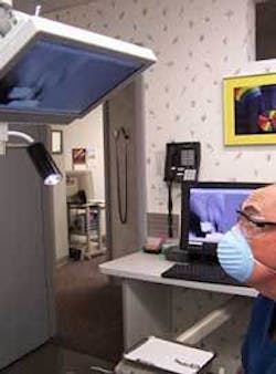
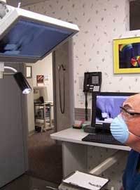
Ideal configuration No. 1 consists of twin LCDs - one behind the patient for clinical management and HIPAA-sensitive data, and one for the patient to view while sitting upright or supine. Variations of ideal configuration No. 1 are often compromised so that utilization, ergonomics, and comfort are sacrificed due to suboptimal placement or makeshift mounting. In this article, the “behind the patient” LCD will be called “BP,” and the patient-viewing LCD will be called “OP.”
Ideal configuration No. 2, for high-tech utilization, adds a third LCD that is more ergonomic and efficient for the clinician to view during treatment. Configuration No. 2 will not be discussed in this article because it is an emerging configuration that is not yet widely used. Viewing clinical data such as X-rays and images on the third LCD requires little, if any, head, neck or eye repositioning during procedures. It is typically located mid-chair at the clinician’s eye level, similar to the LCD location for a surgical video microscope. Pay close attention to the LCD configuration in your operatory. Poorly positioned LCDs can greatly reduce utilization, comfort, and benefits. Optimally positioned LCDs are more ergonomic and reduce fatigue for the operator and patient. When you do it right, patients continually comment that they have a great dental experience.
Digital X-rays
The primary application overlap between the BP and OP LCDs is digital X-rays. Because the LCD becomes your electronic X-ray view box, positioning is critical. The BP LCD is adequate during non-operative periods, but during procedures it involves too much head twisting and eyeball gymnastics. In many cases, the OP LCD works well for viewing X-rays during procedures. Many dentists overlook the importance of LCD positioning when implementing digital X-rays, so be sure to carefully plan where the LCD will be placed in the operatory.
Clinical Research Associates recently published a study that compared the diagnostic quality of digital X-ray systems with standard D-speed film for patients with minimal caries. “When the time was spent to color, enlarge, and enhance the digital X-rays, the diagnostic quality was at least as good as, and sometimes better than, standard D-speed film for some of the digital X-ray systems,” said Dr. Gordon Christensen. Growing acceptance of digital X-rays has been accelerated by the added benefits of digital storage, retrieval, convenience, and reduced radiation.
A number of vendors offer a variety of digital X-ray configurations and benefits. Lightyear Direct (www.lightyeardirect.com), which has more than 3,000 systems installed, offers a two-sensor system with open architecture software that integrates with a variety of practice-management systems. Glen Bachman, CEO, comments, “Digital X-rays offer better diagnostics, improved workflow, and enhanced treatment acceptance at a lower cost than conventional film.” Dexis, which was acquired by Danaher in 2005, has 10,000 installations. “It offers a single-sensor system which is slightly smaller than a No. 2 film that works in conjunction with one universal mount on the screen,” comments Bruce Servine of Dexis.
Suni offers a three-sensor system with a five-year warranty. “Suni sensors are only 3.2 mm thick and provide an easy transition from film to digital,” comments Brian Jaffe, senior vice president of marketing. Digital X-ray systems are also available from Dentrix (www.dentrix.com), Schick (www.schicktech.com), and Kodak (www.kodakdental.com).
Most general dentists are unprepared to invest six figures in 3-D imaging that scans the full oral maxillofacial region in 12 seconds using cone beam technology and provides 150 micron thin image slices, but it won’t deter them from using the images provided by their specialist who has a 3-D system.
“Early next year, Sirona plans to provide viewer software that can be distributed to referring dentists at no charge.” says John Smithson, Sirona’s director of marketing for the imaging group. Sirona provides a range of digital pan/ceph units that can be cost-justified for general dentists as well as specialists.
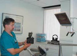
Digital photography
General dentists have finally discovered what orthodontists have known for years - digital photography may be the single most valuable patient motivation tool. Last year digital camera sales exceeded film camera sales, and image quality and value has never been better. Every practice should own and use a quality digital camera. Some companies, like Photomed International (www.photomed.net), for example, configures camera systems specifically for dental applications and provides unlimited phone support for its camera systems. I recommend two cameras for every practice - one a simple, lightweight, point-and-shoot camera that staff can easily learn and use, and the second a higher quality camera for taking high-end clinical photos.
Mike McKenna, a Photomed vice president who has more than 20 years of experience with clinical dental photography, says, “Photomed offers a 30-day money back guarantee. Two popular cameras are the 7.0 megapixel Canon A620 System, which weighs 21 ounces and costs $1,245, and the 8.0 megapixel Canon Rebel XT System, which weighs 53 ounces with the 100mm lens and flash and costs $2,395.”
Orthodontic records have always included a standard series of photographs. I strongly recommend that general dentists have a standard series of patient photographs called an FMI (Full Mouth Images) that is taken for every patient and updated every three to five years.
Another powerful documentation and education tool is a “Panorimage,” taken with a 6.0 megapixel or higher camera and consisting of four simple views - full face, smile (closed bite), upper arch, and lower arch. By using these photos to digitally zoom in and out, patients will not only understand treatment needs, they will clearly understand the precise location in their mouths. With a high-resolution image, you can smoothly zoom from a close-up of a single, unpixelated tooth to a full arch.
Cosmetic simulation
During the past decade there has been broad consensus that simulating patients’ post-treatment smiles is a huge case acceptance tool. Slow acceptance of this technology has been due to the steep learning curve for most software programs, and concerns about meeting patients’ expectations. Practices that have implemented cosmetic simulation have been very pleased with the results. To speed up implementation in your practice, consider using basic features of cosmetic simulation software for simple tasks such as closing diastemas or bleaching.
If hygienists incorporated a three-minute bleaching simulation into every recall visit for patients who would benefit from bleaching, there would be a lot more WOW in the practice, hygienists would become more comfortable using cosmetic simulation, and your practice would bleach a lot more teeth.
Another option is outsourcing cosmetic simulations with companies like Smile-vision (www.smile-vision.net), where you e-mail a patient’s pre-treatment smile and receive a simulated smile by return e-mail to present to your patient.
Patient education
Convenience of the OP LCD is a major factor that contributes to better patient education and treatment acceptance. Applications such as CAESY (www.caesy.com) contain quality images, illustrations, and animations that enhance patients’ understanding and encourage them to accept treatment. Advanced software applications such as DentalImplan provide new tools that dentists and staff can use to present treatment, educate patients, and motivate patients to accept implant treatment.
Dr. Jeffrey Ganeles, DentalImplan’s developer who has an implant and periodontic practice in Boca Raton, Fla., says, “Multimedia tools like DentalImplan provide general dental practices new and simple ways that motivate patients to accept treatment for more complex procedures.”
Entertainment/Distraction
The OP LCDs that patients use to watch TV are becoming as indispensible as anesthetic. Patients are demanding comfortable and relaxing environments where they have some control, even if it only involves changing channels or adjusting the volume. “If my dentist moved, I would go only to a dentist with an OP LCD. It totally transformed my dental visit,” says John Sabina, a representative for Patterson Dental who has been a patient of Dr. John Herzog in Beverly, Mass., for many years. While some dentists feel TV is inappropriate for patients to watch during treatment, most recognize that relaxing patients in this manner reduces stress for both patients and clinicians.
Recent studies have shown that patients who listen to music during surgical procedures require less anesthetic. Major clinical studies have proven that some form of visual stimulation, such as virtual reality programs, are extremely effective at reducing pain for burn patients while surgical dressings are being changed - an excruciatingly painful procedure.
OP LCDs that are properly positioned provide the greatest benefits when used for patient entertainment, with dentists reporting 10 to 30 percent increases in efficiency because patients’ heads do not move, their chins are up, and they lie perfectly still for extended periods of time.
Summary
OP (over the patient) and BP (behind the patient) LCD screens and the applications that use them now provide dentists with powerful tools to provide better dentistry more comfortably and efficiently. A typical investment of $5,000 to $20,000 per operatory in this exciting technology will pay for itself in less than a year, so there is no danger of obsolescence. The investment may seem high to those with no experience with the technology, but knowledgeable users will say it has improved their dentistry, enhanced their lives, and is as indispensible as air and water. Update your practice today, even if you only implement this technology in one operatory. After you gain enough knowledge and experience, you will want to update your other operatories as well.
Steven M. Seltzer is a dental technology expert, Harvard MBA, and practice-management consultant. The inventor of the TLC Technology Lighting Center, has has taught at Boston University, New York University, and Tufts Dental Schools, served as a consultant to the ADA Council on Dental Practice, and lectured at dental meetings for more than 15 years. You may contact Seltzer at (765) 482-9000, steve@hitecdentist,com, orwww.tlcdentist.com


