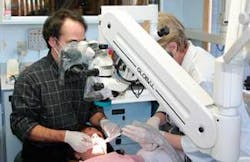
The video-enabled dental operating microscope (VEDOM) with integrated digital documentation capability (DDC): luxury or necessity?
by Glenn van As, BSc, DMD, and Donato Napoletano, DMD
Treating patients in comfort is key to a long and pain-free career in dentistry. Seeing fewer patients and doing more dentistry on each one is key to a highly productive and satisfying career. You canhave it both ways.
The Dental Operating Microscope, or DOM, was introduced to endodontics by Apotheker in 1981, then through further development by pioneers in this discipline, including Carr, Arens, Ruddle, Buchanan, Stropko, and others, the microscope became an important piece in the armamentarium of most endodontists in the late 1990s.
Recent increases in the use and acceptance of the value of magnification in general practice has seen a dramatic increase in the use of medium-powered loupes (3X to 6X magnification) and a growing interest in the use of microscopes for general practice. Microscopes offer four significant advantages in dentistry:
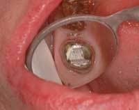
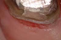
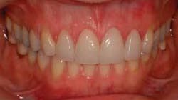
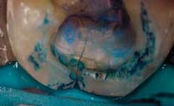
- Enhanced precision through improved visual acuity (Figs.1a and 1b)
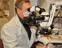
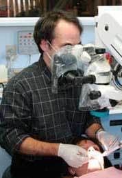
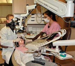
- Improved ergonomics for the operator and assistant (Figs. 2a and 2b)
- Unrivaled ability to document through digital photography and digital microvideography (Fig. 3)
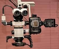
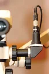
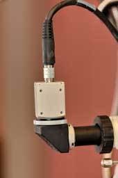
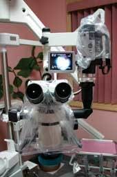
- Microscopes offer a phenomenal opportunity to improve communication with dental auxiliaries, patients, and colleagues through real-time video cameras mounted to the microscope (Figs. 4a to 4c).
Prior to practicing with a DOM, many clinicians consider them to be merely a luxury and that their cost cannot be justified because of a perceived poor return on investment (ROI). The microscope has been used by the authors for between three and nine years, during which time it has become apparent to both of us that the operating microscope is an instrumental part of daily practice. It is utilized for almost all procedures, while also creating a positive ROI for the practice.
The greatest ROI realized for microscope-centered practices are those where the DOMs are equipped with video and digital documentation capabilities and equipped with live video feeds and digital cameras. These microscopes are called video-enabled dental operating microscopes with digital documentation capability, or VEDOM/DDC.
To better illustrate a VEDOM/DDC’s ROI potential, we will start by discussing the benefits of the DOM only, and then discuss how these benefits and the ROI are significantly enhanced when a video source and digital camera are integrated to them.
The DOM offers three main benefits to the operator and patient:
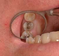
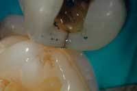
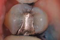
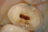
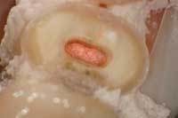
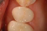
1. Significantly improves visualization via high levels of magnification and unobstructed, coaxial, shadow-free illumination. Increased visualization results in better diagnostic capability, resulting in earlier diagnosis of pathology. Earlier diagnosis leads to earlier treatment and more conservative and faster preparations. The operative time for preparation and restoration is much less for early carious lesions, which results in increased efficiency (especially when treating hard-to-access areas), and hence more predictable treatment outcomes with fewer failures and costly redos (Figs. 5 through 7).
2. Significantly improves ergonomics by practicing in an upright, neutral, and balanced posture looking straight ahead through the binoculars in a comfortable, stress-free position. Operating comfortably means the operator may work for longer periods without suffering from upper or lower back strain or fatigue. This results in increased productivity by doing more dentistry on fewer patients per day. Most patients prefer fewer trips to the office to such an extent that this alone will often stimulate other referrals from happy patients.
3. Significantly decreases the time necessary to successfully integrate various technologies or procedures, such as hard- and soft-tissue lasers, CEREC, and other technologies that help provide patients with minimally invasive procedures. The operating microscope provides the visual acuity that allows for these recent advances to work at peak efficiency. The cornerstone of successful implementation of these technologies is the operating microscope’s unparalleled ability to improve visual acuity. (You can’t treat what you can’t see.)
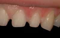
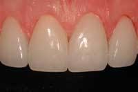
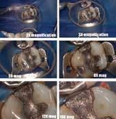
It is easy to see how these first three benefits alone can create significant ROI for the microscope user, but to realize a more significant return, patients have to agree that they have a problem before they can agree to have it treated. This can be accomplished easily and efficiently with a VEDOM/DDC, which enables the operator to share what is visualized in the mouth with the patient. Although some may argue that this also can be done with an intraoral camera, or IOC, the improved picture quality and time savings in obtaining pictures directly through the microscope preoperatively, intraoperatively, and postoperatively make an IOC impractical and inefficient when compared to a VEDOM/DDC. Furthermore, the IOC’s versatility is quite limited when considering that a VEDOM/DDC, with its multiple levels of magnification, can obtain high-quality pictures of not only one or two teeth at medium levels of magnification (5.6X to 9X), but also full smiles, arches, and quadrants at lower magnification levels (2.1X to 4X) and specific parts of teeth at higher magnifications (14X to 21X) (Figs. 8a to 8c). The ability to capture and share this visual information with the patient to demonstrate the need to have dentistry completed is crucial in optimizing the ROI of a DOM to its fullest.
Sharing information with patients to help in acceptance of treatment plans is called co-diagnosis. The VEDOM/DDC allows the clinician to not only co-diagnose but also allows for co-treatment. In sharing the information obtained through a VEDOM/DDC, a few amazing things happen which ultimately result in three additional benefits to the operator, patients, and even staff members. These additional benefits also significantly expand the ROI potential of the DOM alone.
4. Patients suddenly become more interested in what the clinician has to say not just because they, too, can visualize their dental problems better, but because it shows that the dentist cares to communicate better with them; to thoroughly educate them about their problem regardless of its degree of severity. This immediately raises patients’ confidence level and aids in motivating them to treat problems in earlier stages of a disease process, even if it means paying more out of pocket. This results not only in greater treatment acceptance, but also in a significant reduction of time needed to gain that acceptance. Getting patients to accept treatment sooner rather than later also serves to save them money in the long run. The old adage “A picture is worth 1,000 words” comes to mind, but with the microscope, it truly is “a magnified picture that is worth more than words can describe.” In one operator’s practice, when given a choice, 80 percent of patients will view their procedure live at some time during the procedure. Patients who are too apprehensive to watch the live video feed are shown digital images - captured during the appointment - just before they leave the operatory. This process of showing the microscopic images of the completed procedure helps verify the verbal information with visual stimuli. After all, we know that patients only remember 10 percent of what they hear (and only what they want to hear) and 50 percent of what they see.
5.The operator’s ability to document things pre-, intra-, and postoperatively is significantly simplified in photos, which can be obtained in a matter of seconds. This results in more photos taken, not only pre- and postoperatively, but also intraoperatively. Dr. van As routinely takes 20 to 30 photos during a regular 60- to 90-minute procedure. These intraoperative photos are an exceptional means of documentation from a medical-legal standpoint, and they also educate and help patients understand (in very few words and in very little time) why a treatment plan has to be changed suddenly during treatment because of an unforeseen circumstance or complication which can arise during treatment. This is especially important in intraoperative situations where, for example, the clinician discovers a pulp exposure, an asymptomatic cuspal fracture that may result in the need for a crown, additional caries on an adjacent tooth, or a missed canal or complication (perforation) during an endodontic procedure. These photos help communicate why treatment plans and their associated costs may need to be altered. Patients understand such changes much better when visual information is provided. In the 1960s, Mehrabian at UCLA showed that as much as 55 percent of understanding in verbal communication comes from visual cues, and only 7 percent comes from hearing words. Further, 38 percent of the understanding comes from inflections in the tone of the speaker. Using video and/or stills to demonstrate clinical issues helps tremendously to avoid situations where the patient is confused or upset by changes to the treatment plan.
6.Staff members become more interested in what the dentist is doing. With a live video feed connected to a TV monitor, the dental assistant can better visualize and appreciate what the dentist and assistant together are doing to solve a dental problem. By observing a procedure on a 15- to 19-inch LCD monitor in great detail, assistants’ attention and interest levels increase dramatically as they are almost magically transformed from assistants to participants in any procedure. Assistants act as additional sets of eyes for all procedures, and are able to anticipate much better and see much clearer during procedures.
While using an LCD monitor, assistants are able to keep in touch with patients with the macroscopic view through normal vision and, with a quick glance to the screen, they can see the enlarged microscopic view of the treatment site. This ultimately increases assistants’ motivation level, which in turn helps them become more focused and efficient while treating patients (Figs. 9a and 9b).
Although a VEDOM/DDC can raise the cost of a DOM by a few thousand dollars ($2,000 to $6,000 depending on the system), the additional cost is not significant when one considers the added benefits and ROI realized with a video feed and digital camera. The operator should have a positive ROI within the first month or two - generated by increased case acceptance alone - through improved patient communication.
These six benefits occur as the direct result of a properly integrated VEDOM/DDC that is used on almost every patient, including new-patient exams and all treatment procedures. In addition, many other benefits occur as an indirect result of using the VEDOM/DDC, which also add to the ROI. These include increased patient referrals, decreased time required to form a trusting relationship with patients and, perhaps most important, a renewed enthusiasm and higher level of enjoyment in practicing dentistry - just to name a few.
When used appropriately, VEDOM/DDCs can have an enormous positive impact on a practice and practitioner not just financially, but also philosophically, psychologically, physiologically, and emotionally.
Although the authors realize that ROI is an important consideration when purchasing any piece of expensive equipment, we probably never would have acquired three VEDOM/DDCs each if ROI was the determining factor. This is because we really didn’t think they had an ROI associated with them! Our main reasons for acquiring them were purely for the personal convenience of having multiple levels of magnification available at our fingertips (we had become tired of switching between 2.5X and 3.5X loupes), and for improving our quality of work through greatly enhanced levels of magnification. The value of the video and digital documentation capabilities of the operating microscope became apparent afterward, along with positive acceptance from both patients and staff members.
Although many dentists may think they simply cannot afford or justify the cost of a VEDOM/DDC, and that if it came down to choosing between adding another operatory or adding two VEDOM/DDCs (the costs of which are comparable), that the extra operatory would make much more economic sense. Nevertheless, we can only work in so many operatories during the day.
One viewpoint states that having more than three operatories in a solo practice may be counterproductive. It creates a situation that increases stress by running around between ops (typically doing single-tooth dentistry) while decreasing productivity. In contrast, having a VEDOM/DDC not only enables us to significantly increase case acceptance, but also enables us to do more dentistry more comfortably with fewer patients, which can significantly increase productivity. For this reason, we firmly believe that no operatory in a restorative practice should be without one!
In addition, more complex treatments can be attempted when visual acuity is improved, resulting in fewer referrals out of the office, and fewer costly remakes. The focus with microscopes is to use a minimally invasive approach with treatment at an earlier stage. Again, this results in less stress and more productivity. After commanding the learning curve (one to six months), the clinician realizes that greater productivity and increased enjoyment comes with seeing fewer patients and doing more dentistry on each of them.
When considering a VEDOM/DDC for private practice, some clinicians wonder if a personal purchase might not be better. For example, a new car, home remodeling, a long vacation, or a new sailboat might be something the dentist and his or her family consider to be important.
As dentists, we often give our patients the same difficult choices without even realizing it. As we tell them they need three crowns on teeth that are not completely fractured or hurting, they may wonder whether the crowns are really going to be worth sacrificing that new flat-screen TV or family vacation they’ve been saving for.
Your new car or extra operatory won’t help patients make the right choices for them. However, a VEDOM/DDC will! If we really want our patients to make some sacrifices to achieve better oral health, then we might just have to make some sacrifices ourselves.
When you really think about it, patients cannot be expected to agree to treat what they can’t see or understand, especially if they’re not experiencing any symptoms. In addition, we can’t treat anything we can’t diagnose due to restrictions in visualization.
Increased diagnostic capabilities, increased levels of communication, increased precision in treatment, and increased productivity by doing more dentistry comfortably on fewer patients are but a few ways that a VEDOM/DDC realizes its true ROI potential. As the old adage goes, “Seeing is believing,” and creating the microscope-centered practice has been an “eye-opening” experience for both of us. We are sure it can be for you, too!Dr. Glenn A. van As graduated from the faculty of dentistry at the University of British Columbia, Vancouver, in 1987. He immediately joined his father Dr. A.W.H. van As in private practice in Lynn Valley. Since then, Dr. van As has built a high-tech/high-touch practice committed to using the latest technologies to provide the highest level of clinical excellence. Dr. van As has served as an assistant clinical professor at U.B.C., lectured more than 200 times internationally, and is a founding member of the Academy of Microscope Enhanced Dentistry (www.microscopedentistry.com). He may be reached by calling (604) 985-1232 or via e-mail at [email protected].
Dr. Donato Napoletano owns Donato Dental in Middletown, N.Y., where he routinely utilizes technologies such as CEREC, surgical microscopes, and four laser wavelengths in the diagnosis and treatment of caries and periodontal disease. He also owns Donato Dental Systems, a consulting company that helps dentists evaluate, select, and integrate various technologies in a systematic and time-efficient manner. He may be reached at [email protected] or by calling (845) 342-6444.
