Blood-free, laser-assisted granuloma removal and apicoectomy with bone graft
At a time in history, traditional dentistry had a place in all aspects of practice simply because that is what dentists knew. However, it is a new day, and the profession is in a different place. Now everyone is fortunate to have dental technologies at the ready that make everyday procedures—and those we might not have been able to perform before—much easier and more efficient.
One procedure that no longer has to be referred out or looked at as a burden is fibroma removal. With traditional instruments, such as the scalpel, these surgeries are not favored by most dentists. They can be very messy due to blood and are laborious. Additionally, suturing the surgical site adds an extra step and can be difficult. The patient experience also suffers as a result of prolonged time spent in the chair and postoperative discomfort.
Some technologies currently on the market can help erase the stigma surrounding fibroma removals and other tedious soft-tissue surgeries. One is Solea, a 9.3 µm CO2 dental laser that cuts bone, dentin, enamel, and gingiva. Below is a case in which the laser was used during an extensive granuloma removal and apicoectomy with a bone graft procedure.
Case summary
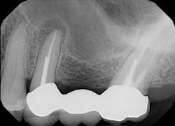
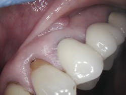
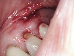
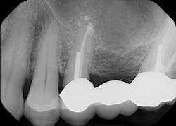
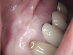
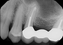
The patient, a 63-year-old female, presented for an emergency visit. She explained her situation, complaining of pain and a lump on the top-left quadrant of her mouth. After an oral evaluation and x-ray (figure 1), it was discovered that she had a large draining granuloma with swelling in the mucobuccal fold near teeth Nos. 12–14 due to a chronic periapical abscess (figure 2).
Clinical objective
The objective was to remove the granuloma at the junction of keratinized and elastic tissue, as well as the periapical pathology, without perforating the sinus on tooth No. 13.
Technique
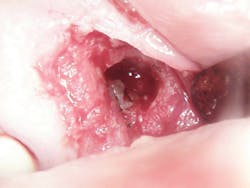
Aiming the laser at the affected area at a 45-degree angle, the first step was to remove the fibroma (figure 3). This was done easily after choosing the appropriate spot size, mist, and power. Following removal, the selections were adjusted for the next step, which was to make an incision in the gingiva. Keep in mind that it was vital to leave approximately 2 mm of keratinized tissue and raise the flap at this time. The laser’s precision allowed for the precise cutting that was needed.
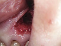
Continuing to adjust spot size, mist, and power as necessary, the laser was used to enlarge the fenestration in the buccal plate (figure 4). The granulation tissue from the abscess then had to be removed in order to expose the root for amputation. Simultaneously, a slot prep was cut for retrograde bioceramic restoration (figure 5).
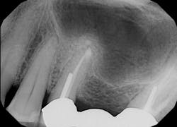
After using a round burnisher and blow test to determine that there was no perforation to the sinus and the membrane was still intact, the laser was used at a spot size of 1.25 mm, on low power mode, and without mist or air to debride the medial superior area of the bony defect. X-rays were taken following immediate placement of bone-graft material to display the site after using Solea to remove bone and granulation tissue (figures 6 and 7). The entire procedure, including the bone graft, took approximately 40 minutes from start to finish.
Advantages of using a 9.3 µm CO2 dental laser
Setting aside the scalpel and using a 9.3 µm CO2 laser for this type of hard- and soft-tissue surgery offers advantages in procedural convenience, safety, and patient experience. In this case, the procedure was performed virtually blood-free, which allowed for a clean site that made treatment simple.
The laser also made ablation of the bony structure easy. If a conventional handpiece or piezo had been used, the cutting would have been nowhere near as seamless or precise. The infected granulation tissue was removed quickly and safely. Another issue that could have occurred was perforation of the patient’s sinus. With traditional instruments, this would have been more likely to happen, causing adverse effects. Using the laser, perforation was not a concern.
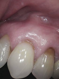
From a patient perspective, a 9.3 µm CO2 laser can dramatically change the dental experience. In this case, chair time was reduced (compare 40 minutes using the laser to the 60–90 minutes required by traditional dentistry), and the patient did not have to worry about excessive blood or postoperative pain. Healing was also rapid without any gingival defects.
During post-op visits, final results at the three- and eight-month marks and x-rays taken at eight months revealed optimal outcomes that met the clinical objective (figures 8–10).
Louis Napolitano, DMD, graduated from Georgetown University in 1977 with a BS in chemistry and earned his doctor of dental medicine degree from Rutgers Dental School in 1982. He completed his general practice residence at Jersey Shore University Hospital and has been in private practice in Jackson, New Jersey, since 1983. Dr. Napolitano has been performing laser dentistry with Solea for four and a half years. He can be contacted at (732) 905-2488 or [email protected].
About the Author
Louis Napolitano, DMD
Louis Napolitano, DMD, graduated from Georgetown University in 1977 with a BS in chemistry and earned his doctor of dental medicine degree from Rutgers Dental School in 1982. He completed his general practice residence at Jersey Shore University Hospital and has been in private practice in Jackson, New Jersey, since 1983. Dr. Napolitano has been performing laser dentistry with Solea for four and a half years. He can be contacted at (732) 905-2488 or [email protected].
