How diagnostic technologies impact your practice and ROI
Dental diagnostics have evolved over the last few years, with new advancements and emerging technology transforming the way doctors and their teams diagnose and treat patients. With so many dental practices expressing growing interest in or having recently adopted newer diagnostic technologies, it can be daunting to figure out how to incorporate these and see a positive impact on your team, patients, and bottom line.
With so much to consider, it’s important to figure out the right time to adopt these technologies by setting strategic goals and researching the resources and education that fit your practice. By taking a comprehensive approach to adding technologies and diagnostic capabilities, dental professionals can experience a greater return on investment (ROI).
When to adopt new technology
Without a clear understanding of the financial and patient care benefits, some dental professionals are reluctant to adopt new technologies. The first step in determining whether your practice is ready for that next piece of equipment or software is to spend time assessing your current strengths and weaknesses and establishing long-term goals. Only then will a strategic plan for investment and implementation of the new technologies become clearer. Before any decision is made, think about this question: “What type of practice do I want in one, five, and 10 years?”
While we have always been big proponents of bringing in new technology, it’s crucial to introduce it wisely. It is unlikely that you will achieve the greatest financial ROI by doing a complete overhaul on all your equipment at one time. Rather, creating a staged onboarding of new equipment unburdens the financial impact as well as eases the learning curve. This allows enough time for training to maximize the ROI and the improvements to patient care.
In 2012, at Downtown Dental, we wanted to set a strategic course for the future of our business. As a result of our planning, we gradually incorporated various equipment and diagnostic tools that built off of each other in order to maximize our ROI and dramatically improve the quality of our patient care. Beginning in 2013, we slowly began to adopt digital dentistry and invested in a hard/soft tissue dental laser (Biolase Waterlase iPlus Er,Cr:YSGG) to offer therapy for surgical, restorative, and periodontal applications. The next year, we moved into CAD/CAM (E4D CAD/CAM System, now a Planmeca product) to fabricate crowns, bridges, and onlays on-site.
In 2015, we recognized the need for clearer imaging and decided to transition into 3-D imaging (Planmeca ProMax 2D/3D unit). That same year, we added implant design modules and more fully utilized the software integration (Romexis, Planmeca).
After spending time mastering our milling system and imaging capabilities, we were then ready to move into the new areas of dentistry that we felt passionate about. We began training in craniofacial pain and temporomandibular joint dysfunction (TMD) with the Las Vegas Institute for Advanced Dental Studies (LVI), and we quickly recognized how imperative and beneficial our current technologies were in these areas of dentistry.
In 2017, we brought in additional new technology, including the BioPak Jaw Tracking and EMG Monitoring System, as well as an occlusal analysis system (T-scan, Tekscan), which enables the monitoring of jaw and muscle movement and micro-occlusion analysis in real time. We use this alongside our 3-D imaging system. Without prior training with our current technology, we would not have been prepared to move into TMD.
Due to how interconnected sleep apnea is with TMD, we saw an opportunity to enhance our practice with sleep medicine. We saw many TMD patients who were also suffering from sleep apnea, so we brought in home sleep tests (MediByte Junior, Braebon) for further diagnostics when our screenings indicated a high risk for obstructive sleep apnea. Our 3-D imaging system has been instrumental in visually showing patients their risk factors for sleep apnea, such as a small waking airway space and a high Mallampati score.
By 2018, we were using all of the technology and equipment we had invested in over the last few years to maximize both our patients’ treatments and the practice’s ROI.
Purchasing criteria
Before a practice commits to new technology, it is important to establish purchasing criteria, focusing on both immediate and long-term goals. While it is true that the “best” technology will be defined differently by different decision-makers, it is essential to agree on the criteria and goals that pertain to your practice as you choose the technology.
The criteria we use to evaluate a company or specific product are training and support, predicted longevity, and the culture of research and development within the company. Support is the most important component you need to achieve maximum ROI.
First, support includes having access to experts who can answer your questions and solve any issues with the equipment or software—not only from the company itself but also from the network of doctors who use the equipment. Second, support includes education and training that will help maximize the value of the technology and the quality of patient care in your practice.
Generally there are two to three heavy hitters for every type of technology, and it is essential to speak with each of them to get a feel for their equipment and the company itself. If the overall goal is growth and advancement of the practice, the company’s R & D history and the product’s ability to work well with any future technology purchases become extremely important. The order of purchasing and integration for the technology should be dictated by your immediate goals, while the company you choose to work with should be dictated by your long-term goals. You must seek to understand how the technology will evolve over time and how it will help your practice in the future.
Addressing integration
You will get the most value out of your new system when your team is properly trained and you have a strategic plan for practice integration in place. Basically, integrating technology comes down to focusing on two essential aspects of educating your team (including the doctors):
- The clinical aspect and how to use the technology on a daily basis
- How to communicate the value of the technology to your patients and how it benefits each patient’s unique desires
With regard to the second component, getting your team on board and making technology part of your practice’s culture are essential for success. Our team can have a diagnostic and treatment conversation with patients because they understand the technology and know how to communicate its value—without us being in the room. So, the ability to train staff effectively and give them the necessary resources are imperative for onboarding and full integration of the technology.
Improved diagnostics and bottom-line implications
Incorporating more diagnostics into your practice not only will allow patients to receive diagnoses quicker and more accurately, but it will also open up additional revenue streams.
With imaging, specifically, there are potential problems that cannot be detected in 2-D imaging x-rays due to limitations such as magnification, distortion, superimposition, and misrepresentation of structures.1 With 3-D imaging, clinicians are able to go through slice after slice and see everything in greater detail, which in turn increases the opportunity for potential treatment.
In our practice, we spent a lot of time learning how to use 3-D imaging, and by doing so, saw an immediate increase of $42,000 in diagnostic revenue during the first year after implementation. This number does not account for the treatment that followed the diagnoses; rather, it is a representation of what a highly educated and engaged team can do. Because they immediately saw the benefits of imaging on patient care, they were better able to advocate to their patients about the importance of timely diagnostics. This is only a portion of how imaging technology can impact a practice’s bottom line, but it demonstrates that education and training are valuable pieces to the puzzle. Imaging allows you to see things that are asymptomatic and not picked up in annual bitewings. Even with abscesses, the only way to catch them fast enough is with 3-D imaging. Resorption lesions can be very difficult to spot with 2-D imaging until it is too late. But 3-D imaging lets you capture the lesions fast enough to potentially save patients’ teeth.
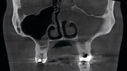
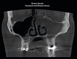
When it comes to sleep dentistry, clinicians can visualize a patient’s airway clearly with 3-D imaging to see if there is a need to test further for sleep apnea. One of the most common things we can see with cone beam computed tomography (CBCT) is sinus infections. We have had many patients who do not realize they have a sinus infection, instead thinking that the pain they are experiencing is caused by something else entirely.
One chronic sinus infection was noted in a scan taken on a patient who was referred to us for sleep apnea treatment. This patient had not been able to breathe out of the left side of his nose for seven years. The entirety of the paranasal sinus, superior nasal passages, ethmoid, sigmoid, and frontal sinuses were involved. Resolution of this pathology and another polysomnogram to evaluate the patient’s obstructive sleep apnea were required prior to initiating oral appliance therapy, as the sinus infection was a major contributor to the patient’s apnea (figure 1).While we have patients who come to see us specifically for our TMD diagnostic offerings and capabilities, others come in for a routine cleaning and talk to us about the pain and symptoms that they don’t know are related to their jaw or bite. Symptoms such as popping and clicking of the jaw joint, chronic neck and upper back pain, migraines, or cranial facial pain can be nebulous, sporadic, and multifactorial. The dental technology we use helps us identify the causes of the pain and understand a complete picture of the patient’s disposition.
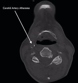
Uncovering incidental findings has had a large impact on our bottom line. Without the right equipment and training, we would not be able to detect or diagnose any of the dental mysteries that are causing patients discomfort. 3-D imaging helps us identify pathology that is unable to be seen on traditional x-rays. And, to some degree, it has made an impact on patients’ lives. We have had three patients in the last couple of years who have had arterial blockages, putting them at a significantly higher risk for stroke. CBCT diagnostics with a large field of view completely turned around the life of one of these patients. We discovered risks for sleep apnea and addressed those; we also uncovered a blockage that, if not addressed, could have dramatically altered the patient’s life.
Because of the adoption of diagnostic technology, our practice is producing 250% ROI from when we first started. There are many contributory factors. For root canals alone, we experienced a $160,000 increase in revenue the year we brought better diagnostics in-house. Since investing in 3-D imaging and becoming proficient at CAD/CAM dentistry, we have produced $1.1 million in additional dentistry for our annual year-over-year growth since integration.
Patient and staff benefits
All things considered, it is important to know that improving diagnostic capabilities for your practice will also benefit your patients. The technology we implemented has allowed our staff to provide more conservative treatment in order to avoid unnecessary procedures. There have also been times when this technology allowed us to be conservative by treating conditions earlier than we would have been able to otherwise.
Bringing in technology means educating your team. A better educated team will have completely different conversations with patients about their dental health. The workflow in treating patients is different because technology increases efficiency. Many of the conversations that patients have are with hygienists and assistants, because they are well versed in using the technology and communicating its advantages. Having a qualified and well-educated team offers patients a different and more pleasurable experience.
From a doctor’s perspective, the most important part to learn for diagnoses is what a normal anatomy looks like. This can make all the difference because you’ll be able to determine what is normal and what needs further diagnostic interpretation. By taking the time to educate yourself and your staff, you will be able to get more accurate visual representations of the clinical problems, which can lead to better patient health and increased revenue.
Conclusion
If you do your homework to find the right diagnostic technology for your practice, create an appropriate plan and execute against it, you will build a larger, more profitable, and ultimately healthier practice.
Reference
- Jain S, Choudhary K, Nagi R, Shukla S, Kaur N, Grover D. New evolution of cone-beam computed tomography in dentistry: combining digital technologies. Imaging Sci Dent. 2019;49(3):179-190. doi:10.5624/isd.2019.49.3.179
About the Author

Daron Clark, DMD
Daron Clark, DMD, a 2007 graduate from the University of Mississippi, is a practicing physiologic dentist and founder of Downtown Dental in Nashville, Tennessee. He is a member of the American Dental Association, International Association of Physiologic Aesthetics, and a fellow at the Las Vegas Institute for Advanced Dental Studies. Dr. Clark serves as a member of the Planmeca Board of Education.

Lisa Smith, MBA
Lisa Smith, MBA, a graduate of Vanderbilt University, is clinical director of Downtown Dental in Nashville, Tennessee. Her role includes strategic planning, vetting new technology, brand development, sales training, improving patient experiences, and removing barriers to better clinical outcomes. She coaches doctors and teams on how to build the practices they want by studying the right business metrics and focusing on communication, education, and technology.


