Handling the emergency referral: the case of the excruciating tooth
For more on this topic, go to www.dentaleconomics.com and search using the following key words: emergency referrals, necrosis, DENTSPLY/Tulsa Dental, Dr. L. Stephen Buchanan.
When your favorite referring dentist calls regardinig a patient emergency, your only answer is, “Send him over now.” Minutes later the guy comes into your office literally staggering with pain. You seat him, take conventional PA X-ray images, gather clinical data, and do just a bit of pulp testing to learn what’s going on. You now know he needs an RCT on tooth No. 19, so you capture a Cone Beam CT 3-D imaging volume and are good to go.
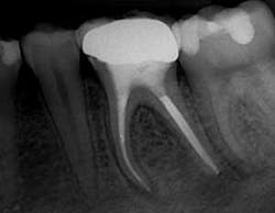
Fig. 1. Post-op radiograph showing severe curvatures of the thin mesial root and the very conservative shape cut by GT Series X Files.
The CT data reveals that there are four canals in this lower molar, that there is a large (nearly the size of a primary canal) isthmus canal between the MB and ML canals at about the middle of the root, and that the distal root has two primary canals that are apically confluent but have an accessory canal bifurcating from the point of confluence.
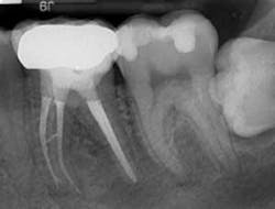
Fig. 2. Distal view showing mid-root isthmus canal and apical bifurcation of the MB canal filled.
This case presents the worst kind of endodontic pain — the dreaded partial necrosis. The coronal half of the pulp is necrotic, and the apical half of the pulp is alive and angry!
These are the teeth that hurt spontaneously, hurt in a prolonged way to heat, or perhaps feel better with something cool or cold on them.
Often you will see patients with this condition in your reception room with an ice-cold can of soda or a cup of ice. The slightest increase in pressure in the dead necrotic pulp space stimulates the vital but degenerating pulp further apical; it hurts to percussion since the disease state has hit the end of the root canal system and is moving through the periapical tissues.
Our emergency treatment plan is for access to be done, the primary canals to be debrided of pulp tissue (the offending organ), and the tooth be left open until I can finish treatment.
We anesthetize the tooth with a Gow-Gates mandibular block with two carpules of 2% lidocaine with 1:50K epinephrine, and a buccal infiltration with one carpule of Septocaine (4% articaine), followed by one carpule of Citanest Forte 1:200K epinephrine given through an X-tip cannula (DENTSPLY/Tulsa Dental).
We accomplished isolation within one minute by using a winged clamp mounted in a prepunched rubber dam stretched onto a circular Nygaard Ostby rubber dam frame, with the dam sealed, using Ultradent’s light-cured blue Blockout Resin.
Access was cut with a No. 4 round diamond bur with water spray, a wet No. 1557 round-ended cross-cut fissure bur, a dry No. 4 round carbide bur to enter the pulp chamber, and an LAX (line angle extension) diamond bur to literally cut 90% of the access cavity in less than two minutes.
Four canals were located with a DG-16 endodontic explorer, and then negotiated in the presence of ProLube lubricant by hand to No. 10K-File and with PathFile .02 taper rotary negotiating files (newly introduced by DENTSPLY/Tulsa Dental).
I cut the .13, .16, and .19 mm tip sizes of PathFiles to length in all of the canals, as determined by a J. Morita Root ZX apex locator.
The procedure was so quick with the rotary negotiation that I realized that this patient, put in my schedule as a walk-in, might actually be completed! The lubricant was hosed out of the access cavity by an air/water syringe, and 17% EDTA solution was introduced as irrigant with a 30-gauge pro-rinse needle.
A 20-.06 GT Series X File (DENTSPLY/Tulsa Dental) was used to cut initial shape in the mesial canals. It hung up about three-quarters of the way down, so I used a 20-.04 GT Series X to cut to length.
An apical irregularity was noted in the MB canal through tactile sense of the files moving to length in that canal. The 20-.06 easily cut to length thereafter, and gauging with nickel titanium K-files (very important to use NiTi here) confirmed apical continuity of taper. The mesial canal shapes were done.
A 30-.08 GT Series X File was cut to length in both of the distal canals. Gauging revealed that the terminal diameter of these canals was greater than .3 mms. So a 40-.08 GT Series X File was cut to length, after which gauging revealed the actual apical diameter of the distal canals to be .55 mms.
Because the distal root was not overly thick, using a 40-.10 GT File (with a 1.25 mm MFD) or a .12 GT accessory file (1.5 mm MFD) would unnecessarily weaken the root structure.
So the 40-.08 GT Series X File with its conservative 1.0 mm maximum flute diameter was taken 2 mm long, thereby cutting a 54-.08 shape and effectively creating apical continuity of shape in a narrow root with a large apical diameter.
I then soaked the tooth in full strength NaOCl for 40 minutes while I finished treatment of my regularly scheduled patient, who had been soaking as well.
Obturation was accomplished with the continuous wave obturation technique. Final radiographs revealed mesial canals with severe curvatures filled with no evidence of apical transportation with an isthmus canal as large as a primary canal cleaned and filled, and an apical bifurcation of the MB canal. The distal canals were apically confluent, and obturated with remarkable accuracy considering the large apical canal diameters.
I called the patient that night, and he reported 90% less pain. One week later, he was 99% comfortable.
L. Stephen Buchanan, DDS, FICD, FACD, is a diplomate of the American Board of Endodontics and an assistant clinical professor at the postgraduate endodontic programs at USC and UCLA. He maintains a private practice limited to endodontics and implant surgery in Santa Barbara, Calif. He is the founder of Dental Education Laboratories, a hands-on training center that serves general dentists and endodontists to upgrade their skills in new endodontic and implant technology. Reach him through Dental Education Laboratories at www.endobuchanan.com.


