Laser-assisted tissue management as the foundation of a digital restorative workflow
Key Highlights
- Shows how predictable soft-tissue management improves scan accuracy, margin visibility, and restoration fit in same-day digital workflows.
- Explains how a 9.3-micron CO₂ laser enables efficient tissue troughing, hemostasis, and preparation without disrupting the digital workflow.
- Demonstrates, through a full-mouth rehabilitation case, how effective tissue control can eliminate temporaries and second appointments while supporting single-visit definitive restorations.
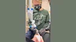
The digital restorative same-day workflow is only as reliable as the tissue environment it depends on. A bleeding margin, inflamed gingiva crowding a preparation, or subgingival decay extending below the tissue crest will compromise scan accuracy and the fit of the final restoration. Managing that environment predictably is, in my experience, the rate-limiting step in same-day digital dentistry.
Our practice in Evansville, Indiana, runs a fully digital infrastructure: intraoral scanning, chairside milling, CBCT, and 3D printing. Over the past six years, the Solea All-Tissue Laser has become the instrument that makes the rest of it function reliably and at maximum efficiency. Operating at 9.3 microns, the CO2 wavelength sits near the absorption peak of hydroxyapatite and water, enabling ablation of enamel, dentin, bone, and gingiva with a single instrument. For soft tissue, it produces reliable hemostasis and eliminates sutures in most cases. For hard tissue, it vaporizes enamel and dentin precisely without the thermal spread that limits other CO2 systems.
Integration with the digital restorative workflow
The weak point in same-day restorative dentistry is the gap between preparation and scan. Retraction cord is effective for straightforward displacement but introduces variability in the presence of inflamed, hypertrophic, or actively bleeding tissue. When tissue control fails at that stage, the case converts to a temporary and a second appointment.
With Solea, troughing is performed after preparation and immediately before scanning. The tissue is incised precisely at the margin, hemostasis is achieved in the same pass, and the field is ready to scan without additional retraction steps. Hypertrophic tissue overlying existing restorations or decay is removed cleanly, allowing preparation to proceed in a controlled field. The result: clean, dry, fully visible margins at scan time.
Clinical case: Full-mouth rehabilitation in a single sedation visit
A patient presented with full-mouth failing dentistry: amalgam restorations with overhangs, secondary caries, and crack lines throughout the posterior dentition. The patient consented to complete rehabilitation under sedation in a single visit.
Following removal of existing restorations, tissue was irritated and actively bleeding at multiple sites, consistent with chronic inflammation from years of microleakage and marginal breakdown, the inflection point at which same-day cases most commonly convert to temporization. Using the 9.3-micron Solea laser, we achieved hemostasis at each site, completed composite buildups where indicated, and finalized preparations across the full arch. We scanned immediately, designed and milled restorations chairside, and delivered permanent crowns before the patient left. No provisionals were placed; no delivery appointment was required (figures 1-4).
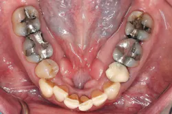
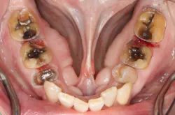
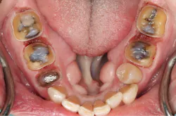
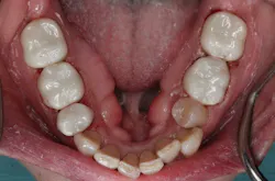
The outcome was a direct function of intraoperative tissue control. Laser-assisted troughing and hemostasis resolved the variables that convert complex cases into multiappointment sequences, allowing the digital workflow to function as designed.
Conclusion
The clinical performance of a digital restorative workflow is dependent on the quality of tissue management at the preparation and scanning stage. In our practice, the Solea laser has addressed that dependency consistently—across routine troughing, hypertrophic tissue removal, subgingival decay removal, and cases complex enough to require osseous reduction before a preparation can begin. Diode and 10.6-micron CO2 lasers are limited to soft tissue and cannot address decay or bone; erbium systems handle both but lack the hemostatic efficiency that makes same-day scanning reliable. The 9.3-micron CO2 wavelength means that whatever tissue obstacle a case presents, the workflow doesn’t stop.
For practices building or expanding digital dentistry capabilities, tissue management instrumentation warrants the same clinical scrutiny as the scanning and milling systems it enables. When tissue is no longer a variable, same-day dentistry becomes predictable and efficient.
Editor's note: This article appeared in the September 2026 print edition of Dental Economics magazine. Dentists in North America are eligible for a complimentary print subscription. Sign up here.
About the Author
 Allie Frounfelter, DDS
Allie Frounfelter, DDS
Allie Frounfelter, DDS, is a leading cosmetic dentist at All in the Family Dental in Evansville, Indiana, known for her transformative smile makeovers powered by cutting-edge digital dentistry. From 3D scanning and Exocad design to 3D printing and precise restorations, Dr. Frounfelter utilizes a fully digital workflow to deliver results that change lives—and smiles—with precision, predictability, and heart. Contact her at [email protected] or on Instagram @dralliefrounfelter.