Ask Dr. Christensen
Gordon J. Christensen, DDS, MSD, PhD
In this monthly feature, Dr. Gordon Christensen addresses the most frequently asked questions from Dental Economics® readers. If you would like to submit a question to Dr. Christensen, please send an e-mail to [email protected].
For more on this topic, go to www.dentaleconomics.com and search using the following key words: methods for crown lengthening, bone recontouring, Dr. Gordon Christensen.
Q: I hear and read about "crown lengthening" in many lectures and publications. I have concluded from most of these encounters that I must lay a flap and remove bone to accomplish crown lengthening. However, I seldom do that procedure, and I do not feel that it is necessary most of the time. Am I missing something important, or should I be doing a flap and bone contouring on a routine basis? I am confused.
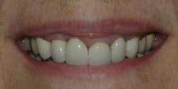
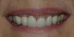
A: Let's see if I can help to clear up some of your confusion. There are many cases that do require bone recontouring. Note the situation in Fig. 1 for which I planned a new fixed prosthesis. If I were to make new restorations for this patient, my restorations would not appear to be any better than the attempt by the previous dentist. The sulcus depth around these teeth is about equal on each tooth.
It is easy to see that removal of just soft tissue could not equalize the gingival levels and still leave at least 1 mm of sulcus depth. Therefore, some removal of bone is mandatory in this case to provide esthetic equalization of the location of the gingival tissues.
Releasing the gingival tissues, removing an appropriate amount of bone, and allowing acceptable healing before making the new restorations is the most desirable therapy in this case.
In the following discussion, I will provide my clinical opinions based on both scientific studies and my own observations over thousands of fixed restorations. Some of my comments may not agree with the comments of others who have written on this subject.
Methods for crown lengthening
There are at least three methods - one used routinely, one used occasionally, and one talked about but seldom used. Let's look at them.
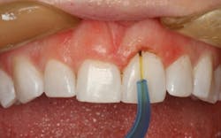
Crown lengthening using a scalpel, electrosurgery, radiosurgery, or laser to simply remove only excess gingival tissue (Fig. 2)
By far, this is the most frequently used form of crown lengthening. In the U.S., almost every dentist has an electrosurgery unit and at least 10% of dentists have a laser. Most practitioners use the mechanical devices to remove unwanted gingival tissues, control blood and other moisture seepage for impressions, and accomplish minor surgical procedures.
The most significant challenge when using this technique is that at least some gingival sulcus depth must be left to allow adequate healing and to produce a predictable healed gingival level. It is common for dentists to leave an inadequate gingival sulcus depth, which often produces irritated and inflamed soft tissue.
At least 1 mm of gingival sulcus depth should remain when using this method of crown lengthening. Assuming that the gingival sulcus depth is 3 mm, up to 2 mm of gingival tissue may be removed and still allow relatively normal healing.
If 3 mm of soft tissue removal is needed for esthetic acceptability, and you remove the 3 mm of gingiva to the level of the bone, the so-called biologic width is "violated." The soft tissue will attempt to grow back but at an unpredictable rate and to an unpredictable level.
The resultant healed gingival appearance is usually inflamed, red/magenta, and unpleasing to patients and you. In some cases, after a significant period of time, the soft tissue may heal, but that healing is unpredictable.
I frequently use the soft tissue only recontouring technique. When respecting the minimum necessary 1 mm of remaining sulcus depth, there is usually no problem with the healed result.
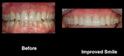
Crown lengthening by removing supporting bone and repositioning the gingival tissues (Fig. 3)
Although a relatively simple procedure, most dentists do not frequently accomplish this procedure. Only long and stable teeth should be considered for this procedure because bone support is reduced in the technique.
Crown lengthening removing bone requires thorough treatment planning and measuring of gingival levels to determine how much removal of bone is necessary to effect an adequate, esthetic gingival appearance when the surgical site is healed.
I suggest having patients look critically at themselves to help you determine the anticipated display of soft tissue and the expected resultant length and appearance of the teeth. A diagnostic wax-up or computer simulation of the anticipated result may be necessary, especially for inexperienced dentists or especially difficult cases.
You should assume that about 3 mm of suprabony tooth anatomy (the biologic width) should be present in the healed state from the new bone level you create to the crest of the gingival tissues. These 3 mm are:
- 1 mm for the gingival crevice
- 1 mm for the junctional epithelium
- 1 mm for the gingival fiber attachment
In an ideal situation, your crown margin will most appropriately be placed halfway into the gingival crevice. I don't know why restorative dentists do not better understand this simple explanation. Perhaps it is the complex nature when it is usually explained in periodontal texts. The summary is - don't expect significantly different results than what I have explained. You can't fool Mother Nature. She wants 3 mm of suprabony tooth structure for adequate biologic width and gingival health.
How long should we restorative dentists wait for optimum healing of the repositioned gingival tissues and optimum bone healing? The longer the better! Any less than six to eight weeks of post-surgery healing is too optimistic.
If you proceed with restorative dentistry before adequate healing and maturation of the gingival position, the plague of crowns will frequently occur - crown margins exposed to the view of observers.
Occasionally, the healed repositioned flap is still not at exactly the right level for an optimum esthetic result. In these situations it is very simple to use electrosurgery, laser, or a scalpel to remove excess tissue from the repositioned flap or adjacent teeth. Obviously, another healing period is required if this is done.
During preparation of multiple teeth for crowns or fixed prostheses, a dilemma frequently occurs. Even though considerable thought has gone into the original treatment plan, caries or old restorations sometimes require that the new restoration margins in the interproximal areas go to bone level or even apical to the bone level. Then a challenge occurs.
Patients expect two appointments for the restorations - a prep appointment and a cementation appointment. They do not expect another appointment after healing for a second tooth prep appointment, for a total of three appointments.
In these situations, removing bone during the crown preparation appointment, providing for the described biologic width, injecting anesthetic with vasoconstrictor into the bone for hemostasis, and making the impression for the crowns will accomplish both the crown lengthening and the tooth preparations at the same time.
Of course, the actual healed location of the interproximal gingival tissues will be unpredictable in such situations, and it is not an optimum technique. However, it is a "real world" occurrence for many posterior tooth situations when the teeth have been mutilated by caries and old restorations.
Orthodontic extrusion to lengthen the clinical crown of the affected tooth
This is a technique that everybody has heard about; however, in my polling of dentists in CE courses, almost nobody does this procedure. Perhaps the delay of restorative treatment and the slight complexity of the procedure discourage practitioners.
If the teeth are long and stable and the patient will accept the additional time involved to extrude the tooth, this is a viable and relatively easy technique. On the negative side, every dentist knows the unpredictability of newly positioned teeth when they have been moved by orthodontic procedures.
Orthodontists often say the teeth need to "sock in" for a significant period of time after active forces have been completed. There is an unknown period of time for the extruded tooth to become stable in the bone, thus not intruding or changing position in the arch.
Further, occlusal forces on the tooth must become neutral for it to be in a stable position. Proceeding before the tooth is mature in the bone is asking for a short crown on the restored tooth.
To summarize my answer, restorative dentists should use crown lengthening on a routine basis. If there is enough gingival tissue present to allow at least 1 mm of gingival sulcus after soft tissue trimming, the removal of only gingival tissue is the best, most predictable, and least traumatic procedure.
In the event that the 1 mm of sulcus cannot remain, bone removal is required. Learn how to do it if you do not know how to do this technique. Infrequently, you may want to consider tooth extrusion, either by you or your orthodontist. Crown lengthening should be a routine part of general dental practice.
Dr. Jon Suzuki, a well-known periodontist, and I, a prosthodontist, just completed a comprehensive, easy-to-understand video on crown lengthening. It shows close-up, live video crown lengthening with bone removal and the subsequent restorative dentistry. It is V4346 "Easy Crown Lengthening." Visit our site at www.pccdental.com or call 800-223-6569 for more information.
Dr. Christensen is a practicing prosthodontist in Provo, Utah. He is the founder and director of Practical Clinical Courses, an international continuing-education organization initiated in 1981 for dental professionals. Dr. Christensen is a cofounder (with his wife, Rella) and senior consultant of CLINICIANS REPORT (formerly Clinical Research Associates), which since 1976 has conducted research in all areas of dentistry.


