Ask Dr. Christensen
Gordon J. Christensen, DDS, MSD, PhD
For more on this topic, go to www.dentaleconomics.com and search using the following key words: cracked teeth, superficial cracks, cracks in dentin, Dr. Gordon Christensen.
Q: One of the most frustrating challenges I see in my practice on a routine basis is cracked teeth, both painful and nonpainful. What is the significance of apparently superficial, nonpainful cracks? I've been told that all obvious cracks should be removed with a bur before restoring the remaining tooth structure. This recommendation seems radical to me, and I've had success when I have not removed the crack. On the other hand, leaving an obvious crack seems too conservative. What do you suggest?
A: Your question is one that each of us practicing dentists faces every day. I will provide some information that I hope will help you make decisions regarding what to do with nonpainful and painful cracks (Figs. 1 and 2).
Superficial cracks (Figs. 3 and 4)
Anyone who has reached middle age will have many visible and nonvisible cracks in their teeth. Banging the teeth together, minor and major accidents, athletic events, and just normal chewing can all cause these cracks. Most of us cannot avoid developing cracks in our teeth. You can easily observe these nonpainful minute cracks by removing your lighted air or electric handpiece from the handpiece cord and allowing the light to come from the cord.
If you place the lighted cord lingual to the anterior teeth, you will see hundreds of cracks in the teeth of a normal adult patient. Most of these cracks are superficial, but if you were to dissect such a tooth, you would see that the cracks usually extend into the dentin. Pain or any other immediate problem condition has not been attributed to these small cracks; however, I see an advantage in showing the cracks to patients to encourage them to reduce their aggressive chewing of ice or other hard and potentially damaging materials.
Treatment of superficial cracks
There is usually no treatment for superficial cracks; however, it is a good idea to educate patients about the presence of these cracks and the potential for them to become worse and eventually break away from the teeth. Most patients do not know that they have small cracks in their teeth, and they're not pleased to know they are there. This information motivates many to reduce or eliminate their aggressive chewing behavior.
As patients enter their mature years, these superficial cracks often become esthetically objectionable. When this occurs, conventional bleaching procedures usually remove the visible stain in the cracks, and superficial "sealers," such as BisCover LV from Bisco or G-Coat Plus from GC America, can be placed on the whitened cracks after a standard acid etching procedure, thus sealing the cracks from more staining for months to a few years.
Cracks into dentin (Figs. 5 and 6)
I'm sure you see these cracks frequently. These patients usually complain of pain during chewing. When you place selective force on the tooth with the well-known Tooth Slooth or similar diagnostic device, moving around the tooth cusps one by one, you elicit pain when applying pressure to one or more cusps. Sometimes a piece of tooth breaks off under the force of the crack-detecting device.
Frequently, the patient calls the office to report pain and make an appointment. Later, the patient often calls back to say that a piece of tooth broke away from the suspect tooth, and there is now no pain. Of course the tooth usually requires some type of restoration, but the pain is gone.
The significance of these cracks is not nearly as important as the cracks described next.
Treatment of cracks into dentin
If the crack is supragingival or subgingival and coronal to the supporting bone, you merely remove the defective piece of cracked tooth and appropriately restore the tooth. The patient is usually comfortable and pleased with the results, and there is no subsequent problem with the tooth.
If the crack extends apical to the bone, this requires making a soft-tissue flap, removing bone to at least 1 mm apical to the crack, allowing bone and soft tissue to heal for a few weeks, and restoring the tooth. This procedure is a typical crown lengthening. If such treatment compromises the esthetic characteristics of the tooth or reduces enough bone to threaten the crown-root ratio, I suggest removing the tooth and placing either a fixed prosthesis on the teeth adjacent to the missing tooth or an implant, abutment, and crown.
Cracks into the pulp or deeply under bone (Figs. 7 and 8)
How do you know if the crack is into the pulp? You don't! However, if the cracked piece does not break away from the tooth when significant force is applied and you cannot see the apical extension of the crack, it must either extend deeply under the bone to an external location or into the pulp. In my opinion, the likelihood of such teeth serving well for an extended period of time is poor.
Treatment of teeth cracked into the pulp or deeply under bone
In my experience, because of the questionable prognosis for such teeth, I prefer to remove the tooth and place an implant if there is adequate bone quantity and quality; however, the patient should be given the option of endodontic therapy and waiting for several weeks after the therapy to determine if the tooth will settle down and become pain-free.
The financial investment for endodontic therapy – potentially placing a post and core, and fabricating and seating a crown – make the decision on these unfortunate cracked teeth an important and expensive one. The overall cost can be $2,000 or more depending on the location of the tooth.
The decision to do this therapy should be made only with a patient's full knowledge of the prognosis, informed consent that the outcome is unpredictable, and knowledge that the tooth may still require extraction. Many patients prefer to extract the tooth and leave the space, have a fixed prosthesis, or have an implant, abutment, and crown.
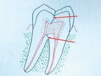
Cracks through the bifurcation or trifurcation of multirooted teeth (Fig. 9).
You've seen patients with a cracked multirooted tooth that segregates the tooth into two pieces. You can determine movement of one portion of the tooth independent of the other portion.
Treatment of segregated roots
Occasionally, such teeth were hemisected, leaving only a single root or two roots of a multirooted tooth (Fig. 10). I have accomplished this procedure many times, but international research on the subject shows limited longevity of such teeth, with an expected service potential of about five years.
In other words, with the impressive longevity of implants now proven to be about 95% at five years, the hemisection procedure that was popular several years ago appears to be less desirable. I suggest placing and restoring an implant in such cases.
A time-saving technique for cracked teeth that you predict are salvageable
I have developed a procedure for diagnosing and treating apparently salvageable cracked teeth that will save time and is relatively simple and predictable. First let's discuss what most dentists do for a cracked tooth.
In a typical office, a patient with an apparently salvageable cracked tooth is treated in the following manner. The technique usually requires a diagnostic appointment, a tooth preparation appointment, and a seating appointment. The patient complains of pain from the apparently cracked tooth. The dentist and staff spend significant time attempting to determine if the tooth is cracked.
Usually, the patient is reappointed for a crown preparation, and finally for a seating of the restoration. The meager revenue from the diagnostic appointment can be avoided by using the following procedure for the initial and subsequent appointment during two appointments instead of three:
Simplified treatment of a salvageable cracked tooth
1. On the first appointment, educate the patient about tooth cracks to allow him or her to decide which treatment will be best.
2. Use a Tooth Slooth or similar device to locate the defective cusp or cusps.
3. Prepare the tooth, make an impression, place a provisional restoration, and seat the provisional restoration with an obtundent cement, usually eugenol. Some noneugenol provisional cements can cause tooth sensitivity after a few days of service, as well as leakage.
4. Call the patient about two days later, after the trauma of preparing the tooth and managing the soft tissue has subsided.
5. Instruct the patient to do the following while the dentist, dental assistant, or front desk person is on the phone: find a wooden pencil, place it on the occlusal surface of the provisional restoration, and bite with significant force on the provisional restoration while rocking side to side and forward and back.
6. Ask the patient if there is pain. If the tooth has a superficial crack into the dentin only, he or she will not feel pain.
7. Assuming the patient does not feel pain, tell your lab to make the crown or onlay.
8. Tell the patient and office staff to make the restoration seating appointment about two weeks later to seat the crown.
9. After desensitizing the tooth with Gluma from Heraeus or a similar product, seat the restoration with a relatively atraumatic, nonsensitizing cement such as RelyX Luting from 3M ESPE or Fuji Cem from GC America.
10. Now, going back to step No. 5, the phone call after the tooth preparation – if the patient still has pain when biting on the wooden pencil, the tooth probably has a crack either into the pulp or deeply under the bone.
11. Appoint the patient for a candid discussion about whether to do endodontic therapy and the needed restorative dentistry or to extract the tooth. Now I prefer to remove the tooth and place an implant with the necessary restorative dentistry. Before implants became mainstream, that would not have been my choice many years ago. Continuing with a root canal and the needed restorative dentistry has many potential reasons for failure on this type of tooth, but I have had success some of the time.
Cracked teeth should not be a mystery. By using the simple step described, you will be able to learn if the tooth is cracked and what to do with it if it appears to be salvageable.
We have at least three videos that will help you with cracked tooth treatment.
Easy Crown Lengthening V4346, Jon B. Suzuki, DDS, PhD, MBA, and Gordon J. Christensen, DDS, MSD, PhD. This presentation shows how to surgically remove bone and reposition the gingival tissues for esthetic reasons or to accommodate removing a crack, as discussed in this article (60 minutes, two hours CE).
Simplified Implant Surgery – The Neoss System V2344, Gordon J. Christensen, DDS, MSD, PhD. This video shows how to place a single implant in a healthy patient with adequate bone. It is essential to solve some of your cracked tooth problems (60 minutes, two hours CE).
Patient Education Video on Cracked Teeth (nine minutes). Your patients will understand the cracked tooth syndrome after watching this brief video, and will understand the need for a crown or onlay.
Call PCC at (800) 223-6569 or visit www.pccdental.com.
Dr. Christensen is a practicing prosthodontist in Provo, Utah. He is the founder and director of Practical Clinical Courses, an international continuing-education organization initiated in 1981 for dental professionals. Dr. Christensen is a cofounder (with his wife, Rella) and senior consultant of CLINICIANS REPORT (formerly Clinical Research Associates), which since 1976 has conducted research in all areas of dentistry.


