Ask Dr. Christensen
Gordon J. Christensen, DDS, MSD, PhD
In this monthly feature, Dr. Gordon Christensen addresses the most frequently asked questions from Dental Economics® readers. If you would like to submit a question to Dr. Christensen, please send an email to [email protected].
For more on this topic, go to www.dentaleconomics.com and search using the following key words: resin-based composite, amalgam, CAD/CAM, Dr. Gordon Christensen.
QI see extravagant claims about the proposed positive properties of composite in Class II locations; however, I have not seen those claims to be true in my practice. Am I doing something wrong or using the wrong brand of composite? Why am I seeing relatively early failure of resin in Class II locations?A A then-major dental company promoted resin-based composite for the first time in 1968. The materials that were available at that time were primitive compared to materials now. In my opinion, the companies prematurely promoted the use of resin in Class II situations in 1968. The materials wore significantly and suffered what has been subsequently called “plucking,” in which the large particles used as filler came loose from the resin, which resulted in microscopic holes in the restoration.Fig. 1: Small, easily restored, two-surface tooth preparation for resin with excellent longevity potential.
In the mid-1980s, resin became more adequate for posterior tooth use, as materials with smaller particle size and less plucking were developed. Additionally, bonding agents became available that reduced the previously prevalent postoperative tooth sensitivity common when the early resins were used in Class II restorations. Dentists and manufacturers began to develop techniques to reduce the occurrence of open contact areas. However, resin still comprised a small amount of the intracoronal posterior tooth restorations placed.
During the intervening years, resin materials and techniques have improved markedly. At this time, resin placed in small posterior tooth preparations serves relatively well. Nevertheless, the research information on the longevity of Class II composites when related to Class II amalgams is embarrassing. There are many controlled research projects that show the longevity of amalgam is about twice that of composite in Class II restorations.
Fig. 3: Moderate-sized, moderately difficult tooth preparation for resin, amalgam, or an indirect inlay requires significantly more time and effort than the preparation in Fig. 2.
My answer to you is not to encourage a return to amalgam
It is my hope you will analyze what the profession is doing wrong to produce such short longevity for resin-based composite restorations.
Recently, the TRAC component of CLINICIANS REPORT (previously called CRA) published a clinical research article showing that the state-of-the art resins are very similar to one another in their ability to provide service in the mouth, and that they have not improved much in many years. I suggest you read the article mentioned above to see the brand comparisons (www.cliniciansreport.org). I am not saying that the resins are inadequate. I am saying that the resins may be only part of the problem for producing shorter restoration longevity.
Tom Limoli Jr. of Limoli and Associates (800-344-2633) has reported third-party payment data to me. According to this data, about two-thirds of the posterior restorations in the U.S. were resin, and only one-third were amalgam during 2010. Only 2% were indirect inlays or onlays.
Currently available radiographic devices, either analog film or digital radiographs, are incapable of showing initial carious lesions. As a result of being unable to see lesions until they become large, dentists make larger, more time consuming, more difficult, and shorter-lasting resin restorations. The following drawings show the various sizes of potential Class II restorations. The time involved, difficulty of placement, and known average revenue for each size is also discussed.
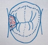
Fig. 1 shows a small Class II tooth preparation entered from the occlusal surface. Some small lesions such as this may also be entered from the facial-proximal surface, if the gingival tissues have receded adequately to allow access for a bur. Often, as shown in the figure, the proximal surfaces of posterior teeth are carious and the occlusal surfaces are not carious.
The small tooth preparation requires only a few minutes, and because of the small size of the tooth preparationFig. 5: Somebody waited too long to restore this tooth, long past the direct placement level and requiring some type of indirect onlay or crown restoration.
This restoration would be designated as an MO posterior tooth restoration for third-party payment benefits. The difficulty for placing resin in this preparation is minimal, and most dentists tell me this is one of the significant indications for resin. The time involvement is minimal for these small preparations, probably about 10 minutes of hands-on time for an average clinician.
The problem with this small restoration is that many lesions that require this type of restoration are missed because of the inability of clinicians to see the lesions on typical radiographs.
Fig. 2 shows a minimally prepared tooth for an MOD restoration. It has been my observation from polling dental CE audiences that most dentists are placing resin instead of amalgam in such small preparations. How much time does such a restoration require for a typical general dentist?
I suggest that a typical experienced practitioner requires only about 20 minutes. However, as with the two-surface restoration previously described, many dentists cannot see such small proximal lesions onFig. 6: Although some dentists heroically restore this type of tooth with direct restorations, a crown is indicated for optimum strength and longevity.
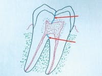
Fig. 3 shows a moderate-sized tooth preparation. What does a typical dentist in the U.S. place in such a preparation? It is likely that most place a resin, but some place an amalgam, and a few place an indirect ceramic or polymer restoration, either made by a laboratory or by an in-office milling device (Fig. 4). What are the challenges if the practitioner waits until a restoration this size is necessary instead of cutting the preparation shown in Fig. 3? The larger restoration requires at least 30 or more minutes in a typical practice, the clinical difficulty is moderate, and there is the chance of making open contacts or leaving occlusion too high. The most significant dentist negatives are that the third-party benefit for this restoration is the same as the much easier and faster restoration shown in Fig. 3.
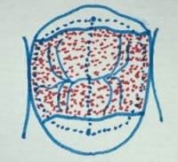
Fig. 5 shows a large MOD restoration. Is a direct restoration indicated in this preparation? In my opinion, the answer is a resounding NO, but many dentists try to use resin in such a situation. I have been criticized for condemning the current generation of large resin restorations.
Such a resin restoration requires 30 to 50 minutes in a typical practice. It is difficult to accomplish, and the likelihood of open contacts, high or low occlusion, over-finishing, and postoperative tooth sensitivity are often high. The frustrating aspects of this large restoration is that the third-party benefit is the same as the amount for those shown in Figs. 3 and 5, the chances of long-term service are very low, and cusp fracture is almost certain after a period of service.
In my opinion, this large restoration should be accomplished only with an onlay or a crown for optimum service, but I recognize well the financial limitations of many patients, and these limitations often lead to inadequate and transitional restorations, doomed to clinical failure.
Fig. 6 shows a classic indication for a full crown. Unfortunately, many dentists attempt to restore such restorative needs with direct materials, usually taking a significant amount of time and providing only short-term, transitional service. The revenue produced by placing a direct restoration in these situations, as dictated by third parties, in no way compensates for the time and effort put into the restoration.
The preceding discussion should make it clear that restoring teeth with direct restorations should be done when the carious lesions are small. This indicates the need for improved methods to detect small interproximal carious lesions. I suggest very careful observation of suspicious proximal tooth surfaces using optimum radiation, trans illumination, and excellent visual observation. Additionally, Kodak provides software called Logicon that has proven to show the nearly exact location and extent of initial carious lesions on proximal surfaces.
When direct resin-based composite is placed in Class II locations in small tooth preparations, the time involved is minimal, the clinical placement is relatively simple, postoperative tooth sensitivity is minimal, the revenue produced is the same as for larger restorations, and the remaining tooth structure retains optimum strength. The clinical longevity of resin restorations placed in small tooth preparations is longer than when placed in large preparations.
The bottom line is that all of us should be detecting carious lesions when they are small, and we should be warning patients of the expected short longevity when tooth preparations are large. In my opinion, having used resin-based composite since its invention, resin serves relatively well in small preps, it will serve for several years in moderate-sized preps as described, and it does not serve well in large, direct-placement intracoronal restorations.
Amalgam use has been dying, but it is still a viable, yet ugly material for those patients willing to accept it. The chances for greater longevity is better with amalgam than with composite in large restorations, but our research over many years shows the longevity of composite to be comparable to amalgam in small tooth preparations.
For detailed information and close-up video of all classes of resin-based composite, see our DVD #V3519, Predictable, Non-Sensitive Resin-Based Composite Restorations. Our course, Successful “Real-World” Practice — Restorative Dentistry also discusses this subject, and attendees receive many samples of the most current, adequately proven products. For additional information, go to www.pccdental.com or call PCC at (800) 223-6569.
Dr. Christensen is a practicing prosthodontist in Provo, Utah. He is the founder and director of Practical Clinical Courses, an international continuing-education organization initiated in 1981 for dental professionals. Dr. Christensen is a cofounder (with his wife, Rella) and senior consultant of CLINICIANS REPORT (formerly Clinical Research Associates).


