A clear view no longer means a stiff neck
The dental-magnifying telescope has become as vital as any dental instrument in providing superior, quality care for the dental patient and life-long occupational health for the dental provider.
Gary A. Morris, DDS, and
Michael I. Kokott, DDS
Long before "windows" became the popular way to visit new worlds with computers, ancient explorers used the telescope as a window to new worlds in the heavens. Today, telescopes also are at the forefront of a revolutionary exploration, opening new windows for dentists to view their clinical world.
Today`s innovative and high-tech optical systems can deliver amazing depths-of-field and wide fields of view that enable the dentist to view a complete oral cavity in focus without having to move. The latest designs in magnifying telescopes, however, can achieve
surprising focus across and into structures of the tooth, as in root canals, for example.
Magnifying telescopes sometimes are called "loupes." A loupe can be as simple as a flat, single-element magnifier (such as plastic magnifying squares that are mounted in front of the eyes) or as complex as a multiple element telescope. Today`s dental telescopes are fashioned from multiple glass lenses that are, themselves, combinations of multiple optical elements. These lenses, called achromats, consist of two or more glass elements, bonded together. A simple loupe typically is inexpensive and lightweight, but can focus at only one discreet working distance. Its poor optical quality, coupled with its limited focusing capability, contributes to eye, neck, and back pain.
High-quality optics also provide a high-resolution image and the ability to discern even the smallest structures - i.e., 50 microns (one-millionth of a meter) and smaller. To maximize the optical performance of the telescopic lens, you should be concerned about the quality of glass used and the basic design of the system. Superior optical systems deliver larger, magnified structures that are not "fuzzy," but are crystal clear. The target image will appear very bright and clarity will be consistent across the entire viewing field, with no distortion or nontrue coloring. In poorer quality systems, image distortion can be seen near the edges of the field (called "vignetting") and chromatic abberations cause yellowing at the edge of the field.
The goal, then, of optimized dental-telescopic design is to maximize resolution, image quality, and width and depth of field, while minimizing size and weight to achieve long-term comfort.
How do these characteristics, once perfectly optimized, combine to affect the practice of dentistry? The answer lies in the realization that 20/20 vision is no longer the state-of-the-art for dental practitioners.
Students and practitioners of all ages are using binocular magnification to improve the quality of their work, increase the efficiency of their practices, and to establish a life-long commitment to a healthful posture. From the moment dentists first use magnifying telescopes, they notice an incredible degree of detail that usually is available only with technical slide presentations. One of the foremost benefits of magnification is in the placement of direct restorations. Magnification offers an instant improvement in the quality of restorative care. The clarity of view enables the dentist to evaluate teeth, excavate caries, refine margins, and place restorations in a precise and efficient manner.
Periodic examinations, as a part of routine prophylaxis visits, are a great opportunity to use magnifying telescopes. Magnification easily allows the dentist to evaluate the margins of restorations, the amount of calculus that needs to be removed, and what radiographs show. Many dentists start out using telescopes for dental treatment only. They soon begin using them into examinations because of their ability to see fine detail. Treatment diagnosis is more thorough, since the dentist can easily detect marginal breakdowns, invisible cracks in both enamel and restorations, porosity in bonding materials, and caries penetration.
Magnification often is an absolute necessity, such as in the evaluation of the passivity of fit in dental implants. The fit of an implant-connecting bar (or multiple implant restorations) is based on clinical judgment. If the interface is equal to the gingival level, or above, the attachment is evaluated visually. This cannot be adequately accomplished without the use of magnification. This also holds true when evaluating crown-and-bridge impressions, as well as the marginal adaptation of definitive restorations.
Periodontists value magnification when treating the trifurcated areas of upper molars and when checking for complete calculus removal. Endodontic procedures that once depended on "feel" now progress by sight with the use of magnification and enhanced, coaxial lighting. This can be quite valuable in increasing the efficiency of previously slow and tedious procedures.
Dentists accustomed to the benefits of magnification frequently use lower-power telescopes for routine, procedures, and then switch to higher-power telescopes for finishing and highly detailed procedures.
Even though the need for magnification transcends the age of the dentist, one of the more obvious demographics that needs optical help is the baby-boomer dentist. This stage of life brings about the advent of presbyopia, a condition that causes the lens of the eye to stiffen and resist focusing at near distances. Although bifocals will correct presbyopia for reading, many dentists also turn to their optometrist or ophthalmologist to provide "enhanced" bifocals for clinical procedures. However, binocular magnification, with its substantial depth-of-field, is the only solution to this problem that will not cause eyestrain and ergonomic difficulties. Maintaining a focus at one discreet distance during procedures that require varied angles of approach is a sure recipe for eye, neck, and back strain.
Binocular magnification allows the practitioner to sit fully erect, maintaining excellent posture, yet still see the operating site with perfect acuity. This means that students and younger dentists now have an edge that their predecessors did not. By establishing good posture habits early in their careers, they will avoid the pinched nerves, bad backs, migraine headaches, shoulder pain, stiff necks, and numbed extremities that some dentists now experience on their way to shortened careers.
Aside from enabling the operating site to be clearly viewed at a distance, how do magnifying telescopes promote good posture? The key to good posture depends on the degree to which the dentist`s head is directed downward while viewing the operating site. Too many dentists spend their entire workday with their head and neck tilted at decidedly unnatural angles. Binocular telescopes can be aimed at comfortable working angles in various mounting schemes to achieve good posture.
No discussion of dental magnification would be complete without considering the role that light intensity and light position play in the performance of the telescope. Maximum resolution and depth-of-field is achieved in any optical system by providing an intense light that is beamed directly down the viewer`s line of sight. Lighting such as this is described as being "coaxial." Coaxial light illuminates the target site without shadowing, and it maximizes the effect of the light`s intensity on the performance of the optical design.A truly coaxial light, coupled with an optical system that delivers a long depth-of-field, can enable the dentist to see clearly and in-focus into all orifices, including the apex of root canals.
For lighting to be coaxial, it must be mounted between the viewer`s eyes (or telescopes), but it shouldn`t obstruct the field of vision. An efficient method for accomplishing this is to mount the light to the eyeglass frame or to the telescope hinge. An important consideration, then, is to minimize the weight of the light to preserve the wearer`s comfort.
Two types of lights are used to accomplish coordinated lighting with dental magnifying telescopes: electric and fiber-optic. The electric dental light has a bulb supplied by an electric power cord. The bulb is contained in the housing mounted to the dentist`s glasses. On the other hand, the fiber-optic light consists of a housing with one or more optical elements supplied by a fiber-optic cable. The light source for the fiber-optic unit is located in a remote electrical appliance. Light is transmitted through the fiber-optic cable to the lens assembly worn on the user`s face.
Fiber-optic lights typically are much brighter than electric lights. This is due to the limited size and intensity of the bulb that can be used. Larger-wattage bulbs placed close to the wearer`s face are hot and uncomfortable. Because fiber-optic lights are fed by remote light sources, the heat generated is isolated away from the user`s face. This design advantage means a light of much greater intensity can be generated without consideration to heat and added weight.
The science of astronomical-telescope design has progressed since the time of Galileo to produce the largest telescope in history - the giant, 10-meter, Keck-reflecting telescope at the Mauna Kea Observatory in Hawaii. Similarly, dental-telescope design has advan-ced to provide lightweight optical systems that deliver high-resolution images with expansive depths- and widths-of-field. Newer dental telescopes with adjustable focal lengths and titanium-mounting systems are pushing the edge of traditional limits at higher levels of magnification.
The dental profession has recognized that 20/20 vision is no longer adequate to meet the challenges of today`s materials and techniques. The dental-magnifying telescope has become as vital as any dental instrument in providing superior, quality care for the dental patient and life-long occupational health for the dental practitioner.
Types of telescope mountings
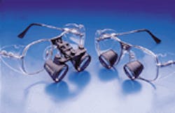
Magnifying telescopes are mounted to either spectacle frames or on a headband. The type of telescope mounting is either on a flip-up hinge or glued directly into the frame`s eyeglass lens. The latter method is typically called "through-the-lens" or TTL.
Performance trade-offs exist between flip-up style, hinge-mounted telescopes ("flip-ups"), and TTL-mounted telescopes. These trade-offs have nothing to do with the optical performance of the telescope design, but are associated with comfort, convenience, and the relationship of the telescope to the wearer`s eyes.
Flip-up telescopes are considered by many dentists to facilitate the "up and down" style of today`s hectic practice schedule. If designed properly, the busy dentist can wear these telescopes all day, flipping them up when not in use.
TTL telescopes, however, must be removed when not in use because the telescope barrels, mounted in the eyeglass lens, may obstruct the wearer`s vision. This is particularly true if the dentist requires a prescription lens to correct for a nearsighted or farsighted condition and/or astigmatism, if he or she needs reading glasses, or a combination of these aids are necessary. A dentist who requires a prescription lens may have to remove TTL telescopes and replace them with prescription glasses between patients.
On the other hand, TTL-mounted telescopes are quite popular. The reason for their popularity stems from the inherent comfort and improved field width that is produced by mounting the telescopes "through-the-lens."
When the telescopes are mounted in the eyeglass lens, they are positioned closer to the wearer`s face and eyes than the flip-up style. This favorable juxtaposition reduces the apparent weight felt on the wearer`s nose.
Because the telescopes are nearer the eyes, the width of field is increased. Some dentists willingly sacrifice the overall convenience of flip-ups for the reduction in weight and increased field width of TTL telescopes.
It should be noted, however, that the differences between the two mounting methods are not extreme. Since both styles serve dentists well, the decision to wear one or the other is reduced to a matter of personal preference.
Examples of two mounting methods: Flip-up telescopes on the left and TTL (through-the lens) telescopes on the right.

