Cone beam: past, present, and future
For more on this topic, go to www.dentaleconomics.com and search using the following key words: cone beam, 3–D imaging, CBCT, digital imaging, Dr. Terry L. Myers.
Here's some good news to start the New Year. Based on U.S. Internet searches, Google's chief economist is cautiously optimistic that the economy is in recovery mode. There are fewer searches regarding unemployment benefits, and more for homes and real estate agents. Economists are predicting that federal funding will create more jobs in the public sector, and tax incentives will encourage expansion in private enterprise. While we understand that recovery will take time, this gives us time to ensure our practices are running efficiently, effectively, and affordably to welcome back the established and new patients who soon will have the funds to renew their commitment to dental health.
Now is the time to make technology count, clinically and financially. Cone beam imaging technology is one of the more efficient and effective ways to improve your clinical skills and increase your business.
The history → CBCT technology dates back to the development of X–ray computerized tomography (CT) in the 1960s. Godfrey Hounsfield won the Nobel Prize in 1972 in medicine for pursuing the first clinical studies utilizing this breakthrough technology. In 1998, the ability to acquire volume (CBCT) data entered the dental realm. Today, cone beam imaging is growing in popularity for dental practices because of its characteristic low radiation, high spatial resolution, and relative low cost (to practitioner and patient) compared to conventional hospital CT scans.
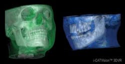
The technology → Compared to fan–shaped CT beams, CBCT images are captured by a cone–shaped beam (hence the name). The beam is emitted in short pulses with a single 180–degree rotation, thereby decreasing the overall radiation exposure — up to 10 times less radiation than traditional CT scans. Unlike time–consuming hospital machines, the in–office cone beam is captured in as few as 8.9 seconds, and reconstructed in less than 20 seconds. The final result is a three–dimensional image that can be sliced and rotated for diagnostics and treatment planning, determining precise tooth positions, bone dimension and quality, TMJ disorders, and other dental conditions. Cone beam technology shows its flexibility with adjustable collimation allowing for image sizes that suit individual needs of the patient and dentist. Scan sizes range from single–arch view up to cephalometric. My system (Gendex GXCB–500™, powered by i–CAT®) has four scan sizes and also takes a 2–D pan. Groundbreaking sensor technology, such as an amorphous silicon sensor, allows for the clearest data capture, with no artifacts or blurriness. My included software is easy to use, compatible with other 3–D imaging applications, and simple to share among colleagues.
The economy → 3–D images allow us to know more about patients' anatomy, giving us greater treatment predictability. The information I can obtain from my cone beam machine has given me the confidence to expand my practice to include procedures, such as implants. My patients are better informed about the costs of total treatment because I know so many details — no surprises or guesswork discovered during surgery. Also, after I educate patients with their 3–D scans, they are more inclined to accept my treatment plans. My cone beam system has directly increased production from added procedures and scan fees. This is not a technology of the future; it is technology that is much needed and available in the present. Cone beam imaging affects every aspect of my practice — from diagnosis to treatment to my financial bottom line. As the economy improves and people recover funds to pursue their dental goals, 3–D imaging has become an integral part of my economic stimulus plan.
Dr. Terry L. Myers has a private practice in Belton, Mo. Contact him by e–mail at office@keystone–dentistry.com.

