Digital Radiography in Pediatric and Special Care Dentistry
I have had the pleasure of working with digital radiography for the past six years. Most of us are familiar with the benefits this technology can bring to any practice, including higher quality radiographs than are possible with film and tremendous efficiency gains. In this article I will explain the benefits specific to pediatric practices and to practitioners who work in hospital operating rooms.
One of the major benefits of digital radiography is patient safety, with a strong emphasis on making sure that children are not exposed to high radiation doses. Next is patient education, which includes not only making young patients feel more comfortable, but also ensuring that their parents understand the recommended treatment. Digital X-rays have greatly increased our ability to respond rapidly to patients’ traumatic injuries, where quick action often means the difference between saving and losing a tooth that has been dislocated or avulsed in an accident or sports competition.
I know that all dentists, no matter what their specialty, are concerned with their patients’ well-being. Where children and radiation are concerned, I am convinced that it is our responsibility to do everything in our power to reduce their exposure. The fact is, the younger the child, the more potential of damage from excessive radiation.
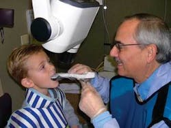
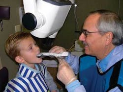
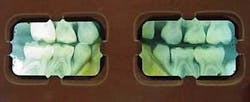
Using digital radiography may reduce the patients’ exposureto radiation. Even if this were the only benefit I realized by switching to digital, I would not have hesitated to install my digital X-ray system. But improving patient safety through reduced radiation is not the only benefit of digital radiography in a pediatric practice. When you’re working with children and adolescents, you’re really working with their parents as well, and this means that patient education and treatment acceptance issues can often be compounded. I found that our conversion to digital resolved many of those issues in a very positive way (Figure 1, above, and Figure 2, top).
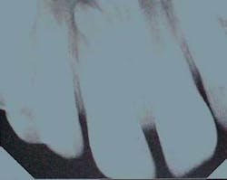
Among other things, I take advantage of the ways digital X-ray images can be manipulated and presented to get children involved in and comfortable with their dental treatment. First, kids think digital X-rays are “really cool,” to quote several of my young patients. They love the fact that they’re seeing large, clear images of what’s inside their teeth, and they love it when I increase the contrast or use color to make things even clearer (Figure 3).
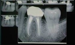
The truth is, their parents love it, too. And that’s the second patient education benefit my digital radiography system brings to me. Parents are much more accepting of my treatment recommendations, because they can see very clearly any caries or lesions or cracks in their children’s teeth. The fact that their children are engrossed in “helping” me with the diagnosis doesn’t hurt their acceptance either. Speaking of diagnosis, we have all had the experience of our patients’ X-rays deteriorating over time (Figure 4). This does not happen with digital X-rays.
Digital X-rays are also an excellent educational tool in another way. If my young patients happen to be studying teeth at school, I can print out images of their X-rays (regular copy paper is excellent for this) so they can take them to school for show-and-tell. They love it!
The area where the benefits of digital radiography’s instant-image capability are perhaps most important is in dealing with traumatic injuries. Here, time is of the essence, and digital X-rays definitely save time!
Recently, I treated a 10-year-old who had had an accident on a school field trip to downtown Chicago. Justin had stumbled and knocked out his upper right central incisor when he fell against one of Chicago’s most famous landmarks, the Picasso statue in Civic Center Plaza. His teacher had the presence of mind to place the tooth in milk, and Justin was rushed to my office, arriving about 50 minutes after the accident. His parents had been notified, and they arrived at my office just before their son.
Both Justin and his parents were upset about the incident, and I quickly took a digital X-ray, as I do in all such cases. I didn’t have to wait for my assistant to develop film; the image appeared immediately on the large-screen monitor in the operatory. As it turned out, Justin had squirmed just as the X-ray was being taken, so I had to take a second one. Because I had to take two X-rays, the time saved over film was as much as nine minutes or longer. This was time that was very important in making sure that the reimplantation of Justin’s tooth was successful. Justin’s parents realized that the time saved through digital X-rays increased the likelihood that their son’s tooth would be successfully reimplanted. (I’m happy to report that Justin’s tooth was saved.)
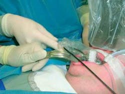
Beyond its importance as a time-saver in trauma cases, digital radiography is very helpful in the operating room. Instead of waiting for X-rays to be developed, we can begin the dentistry right away. This saves the patient from being under general anesthesia any longer than necessary. Also, if you want to check an implant or extraction site or endodontic procedure, you have the benefit of viewing the radiograph immediately. Take your digital X-ray sensor and laptop computer into the operating room. You can store your images and transfer them onto your office computer’s hard drive when you return to the office (Figure 5).
Finally, orthodontists have sent me hard copies of their digital radiographs showing the teeth they would like for me to extract on our mutual patients. This saves time for the dental team and the cost of duplication. There have been several instances in which parents forgot to bring the radiograph and I called the patient’s orthodontic office to e-mail the radiograph to me. This saved time and money in not having to either retake the radiograph or reschedule the patient.
Take a careful look at the various digital radiography systems on the market to see which best fits your practice. You’ll be glad you did.Dr. Fred Margolis is in the full-time private practice of pediatric dentistry in Buffalo Grove, Ill. He is a clinical instructor at Loyola University’s Oral Health Center, and director of the Institute for Advanced Dental Education. He is a fellow of the Pierre Fauchard Academy, International College of Dentists, American College of Dentists, and the Odontographic Society. He can be reached at [email protected], or call (847) 537-7695.
