An eye-opening experience
By Neal Patel, DDS
I have a passion for technology because it bridges the gap of communication between clinician and patient. But let's be honest. Sometimes clinicians cannot agree on a diagnosis for a given patient. As dentists, we are responsible for providing an accurate diagnosis first and foremost. The treatment that is derived from this diagnosis defines us. Why can't we agree on a diagnosis? It is likely due to the fact that we are all using different imaging modalities when it comes to our intraoral X-rays. To this day, there are clinicians who provide film-based imaging for their patients … you know who you are, so don't skip this article. I am sure the sales reps have already worn down the knocker on your door to get you to switch, but for some reason you have not. So, for those of you who have decided to stay with film (for any reason), this article is dedicated to you.
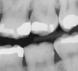
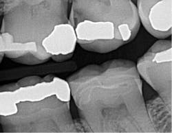
There is a direct ROI when using this sensor compared to older digital sensors or even film. I can appreciate a significant improvement in identifying interproximal and recurrent caries on patients that I thought were completely healthy. It honestly is scary what I had been missing with the previous generation of digital sensors. I also like Schick 33's presets called clinical-task-specific mapping. I can click on a preset and images automatically default to the setting I need -- general dentistry, endodontics, periodontics, or restorative dentistry. Once the image is captured, I can instantly adjust sharpness by moving my cursor left or right over the graphical slider.
Prior to digital sensors, I often would inform patients that "I am not sure how deep the cavity is, but once we start the recommended procedure, I will be able to assess the situation.” Today, with the clarity provided by this sensor, I find myself having the opportunity to explain my findings more objectively to my patients. As a general dentist, I feel responsible for providing all my patients the best comprehensive exam possible. This sensor allows me to diagnose my patients more confidently, and with the help of the imaging software tools, helps me to better explain my findings with greater ease.
The increased confidence in my interpretation of 2-D images from Schick 33 has directly affected my patients' acceptance of their treatment needs. My staff is overwhelmed by our ability to see consistently crisp and detailed digital images. With the combination of digital radiography's low-dose radiation and super crisp images, we do not hesitate to perform X-rays on all patients, and we value the diagnostic details that were once left behind.
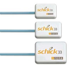
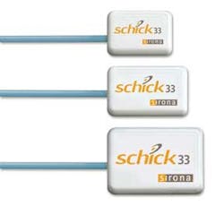
We use all three sensor sizes (0, 1, and 2) in my practice. If we have a patient with special positioning needs, we also can take advantage of the different cable lengths (three, six, and nine feet) and switch them out quickly. Schick 33 has opened my eyes to newfound pathology and restorative needs for many of my patients. My experience has been enlightening, and I treat all existing patients as new patients during their routine exams and cleanings. Perhaps most importantly, support and training are essential, and both Patterson and Sirona rolled out the red carpet with support at the right caliber for the product.
______________________________
Other articles by Dr. Neal Patel:
______________________________
Neal Patel, DDS, created a completely digital practice where he utilizes digital technology throughout (all-digital planning, fabrication of splints, surgical guides, and prosthetics), bypassing traditional analog methods. Widely published, he is best known as an international educator on 3-D digital imaging, treatment planning, and computer-guided implant surgery. He can be reached via email at [email protected].
Past DE Issue


