Smile enhancement using all-ceramic crowns and veneers
Case presentation by Ross W. Nash, DDS
Practice management considerations by Debra Engelhardt-Nash
A young woman presented to my office requesting smile enhancement. Another dentist had previously placed all-ceramic crowns on the maxillary central and lateral incisors. They had been in place for a number of years and had served her well.
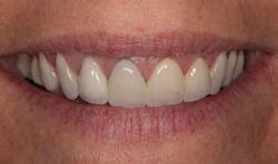
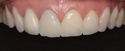
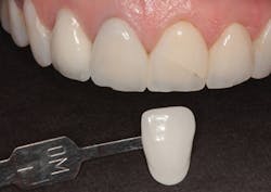
In Figure 1, you can see a close-up view of her smile as it was when she presented. She recently had noticed a fracture in both central incisor crowns and, since replacement was necessary, wanted to investigate the possibilities for improvement. The retracted facial view can be seen in Figure 2. Clinical and radiographic examination showed me that her overall dental health was good, with no periodontal disease or active caries. The patient expressed an interest in widening her smile and achieving a lighter color. The shade she desired was OM1 on the Vita Toothguide 3D-Master shade guide (figure 3).
Figure 1: Close-up view of the patient’s smile before treatment
Figure 2: Retracted facial view of the patient’s smile before treatment
Figure 3: The patient’s desired shade compared to her initial smile
Treatment plan
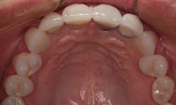
In the occlusal view in Figure 4, notice that her premolars are slightly rotated. Since both premolars were visible in her smile, it was decided to add porcelain veneers to the canines and premolars. The veneers would provide the appearance of well-aligned premolars, as well as add a lighter color. We both agreed that 10 restorations from second premolar to second premolar would be ideal.
Preoperative appointment
At the preoperative appointment, a facebow record was taken (Denar, Whip Mix) and an accurate occlusal registration was made (Futar, Kettenbach). Maxillary and mandibular impressions were taken with an A-silicone alginate substitute material (Silginat, Kettenbach). These were sent to the dental laboratory (Frontier Dental Laboratory) for mounting and wax-up of the upper model and fabrication of a lab putty stent for temporization.
Preparation appointment
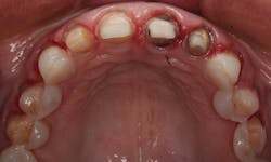
The old crowns were removed; the preparations for the maxillary lateral and central incisors were refined. Veneer preparations were completed for the canines and premolars. In Figure 5, you can see the preparations for four all-ceramic crowns and six veneers. Note that even though there were slight rotations, it was not necessary to break the contacts between the bicuspids and cuspids. This allowed for a more conservative approach that was less invasive. The prepared teeth are shown from the facial view in Figure 6. A new occlusal registration was made using Futar.
Figure 4: Occlusal view of the patient’s initial smile
Figure 5: Occlusal view of the prepared teeth
Figure 6: Facial view of the prepared teeth
Final impressions were taken for the maxillary arch using an A-silicone material (Panasil, Kettenbach). Provisional restorations were fabricated using Visalys (Kettenbach)in the lab stent made over the wax-up, and the case was sent to the laboratory for fabrication of the crowns and veneers.
Practice management considerations: The right way to suggest smile enhancement to patients
Often, new and existing patients come to us with dentistry completed in previous years. Techniques and materials may have dramatically changed since the original restorations were placed. Think about home appliances: Have they added new features over the past two to five years? And what about automobiles or smartphones? The standard or even state-of-the-art care 5, 10, or 15 years ago may have undergone dramatic advancements.
Is there potential for improvement? Possibly. Probably. But how do you approach the subject? And when? And who does this? Can it be done without offending the patient or denigrating the previous dentist? Absolutely.
How? The patient may not initiate the conversation, but may be intrigued to pursue one if given the opportunity. As a consultant, I visit several offices annually. I see dentists—in the hustle and bustle of staying on a treatment schedule—miss obvious opportunities to bring up dental improvements. Begin all appointments with a conversation, not a procedure. Establish a relationship before you establish a procedure code. Codiscover treatment possibilities. Consumers “buy” from people they like—people with whom they have relationship. Plan time in the appointment regime to talk. Most patients are visual learners; they have to see to understand. Use visual aids such as photography of similar cases, video learning, and treatment simulation to help the patient see what you see.
When? The more you incorporate conversations into the routine of new-patient and recare examinations, the simpler the routine becomes. Make it the standard, with few exceptions, to introduce smile rejuvenation or enhancements to your patients. Let them decide whether or not they are candidates for the beautiful dentistry you can provide. Never assume patients cannot afford or aren’t interested in improving their dental health or appearance.
Who? The opportunity to present treatment enhancements begins with the practice’s website and social media platforms, but it must be carried through by everyone on the team. From the initial visit to the recare appointment, everyone on the team is responsible for highlighting treatment opportunities that exist in the practice. With training and practice, it can be done with professionalism and consideration that will put patients’ interests first.
—Debra Engelhardt-Nash
Laboratory fabrication
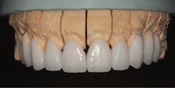
At the laboratory, the model was poured and the model of the maxillary prepared teeth was “jumped” to the previously mounted lower model using the new occlusal registration. IPS e.max (Ivoclar Vivadent) crowns and veneers were fabricated with an HTBL1 (highly translucent) ingot using the lost-wax technique. After the case was pressed, all restorations were cut back and layered to achieve maximum esthetics. They were etched with hydrofluoric acid. The final restorations can be seen on the working model in Figure 7. They are shown photographed on a mirror surface in Figure 8.
Figure 7: Final restorations on the working model
Figure 8: Final restorations on a mirrored surface
Placement
At the placement appointment, the provisional restorations were removed and the IPS e.max restorations were tried in and approved by the patient. They were bonded in place by acid etching the enamel of the teeth prepared for veneers for 10 seconds with an etching gel (Etch-37, Bisco) and liberally applying a universal bonding agent (All-Bond Universal, Bisco). The bonding agent was thinned with air from an air/water syringe and light cured for 10 seconds with an LED curing light. A light-curing resin cement (eCement, Bisco) was placed on the intaglio surfaces of the veneers; they were then placed on the prepared teeth and cured with an LED light. The dual-curing version of the eCement was used for the four crowns after the universal bonding agent was applied to the prepared teeth and light cured. The occlusion was adjusted where needed with fine finishing diamond burs (Brasseler) followed by polishing with porcelain polishing cups (Brasseler).
The final result
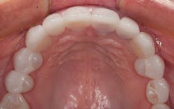
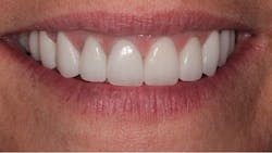
The final result can be seen from the occlusal view in Figure 9 and the retracted facial view in Figure 10. The patient’s new smile can be seen in Figure 11. Utilizing IPS e.max crowns and veneers, we were able to enhance the patient’s smile by creating a lighter color and a wider, more attractive look.
Figure 9: Occlusal view of the final restoration
Figure 10: Retracted facial view of the final restoration
Figure 11: The patient’s new smile
Author’s note: Dr. Nash would like to thank the expert ceramists at Frontier Dental Laboratory for fabrication of the restorations in this case.
Ross W. Nash, DDS, maintains a private practice in Huntersville, North Carolina, where he focuses on esthetic and cosmetic dental treatment. He is an accredited fellow in the American Academy of Cosmetic Dentistry. Dr. Nash lectures internationally on subjects in esthetic dentistry and has authored chapters in two dental textbooks. He is cofounder of the Nash Institute for Dental Learning in Huntersville, North Carolina.
Debra Engelhardt-Nash is a trainer, author, presenter, and consultant, and has presented workshops, nationally and internationally. She is currently the president of the National Academy of Dental Management Consultants, where she is a founding member. She is on the board of the American Academy of Dental Practice. She was a contributing editor for Contemporary Esthetics and Restorative Practice magazine, Contemporary Dental Assistant magazine, and has written for a number of dental publications.

