With comprehensive imaging technologies, patient education leads to practice benefits
Gary Tomack, DDS, FAGD
Many technologies on the market today have raised the bar for dental care. While each has its benefits, certain imaging technologies used as a part of a comprehensive protocol can bring advantages to all involved—from the dentist, to the patient, to the clerical staff. In this article, I will discuss my experience of how digital x-rays, caries detection devices, and intraoral cameras can together benefit patients and the dental practice.
Digital radiographs have had a far-reaching effect on my practice. Before we switched to a digital sensor (DEXIS Platinum), using traditional film radiography was a chore. X-ray film had to be purchased, and it had to be processed in a machine using hazardous chemicals. These chemicals needed to be stored and disposed of properly. Years ago, these chemicals could be poured down the sink, but in recent years, we’ve become more aware of the environmental consequences of hazardous waste disposal. Today, disposal of these products requires a hazardous waste carter. To summarize, using film x-rays was costly both in money and space. Moreover, after taking a film x-ray and waiting for it to be developed, if the image was not good enough, then the whole process had to be started again.
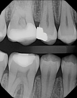
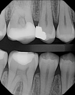
DX Platinum VBW x-ray
Digital x-rays brought us instant images and used no chemicals or film. They eliminated costs and messes. Digital images now provide an interface between me and my patients. I can instantly display images on a 32-inch screen above the chair for patients to see. In my opinion, this instant view is invaluable when doing implants or endodontics. It’s a substantial time-saver. I can enlarge, manipulate, colorize, and draw on these images, which contributes to patients’ understanding of their dental problems. Some patients don’t want to know their problems! But if I can see the problems clearly, I also can identify an appropriate solution and communicate it to them. This type of involvement can lead patients to accepting treatment that is in their best interest and ultimately helps build my reputation as a responsible, caring dentist.
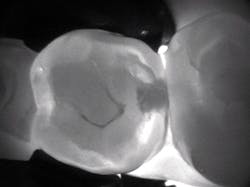
DEXIS CariVu image
In my practice, a caries detection device provides an excellent adjunct to radiographs. We use a device (CariVu, DEXIS) with near-infrared transillumination technology to produce images that look similar to x-rays. For example, a potential Class II lesion may appear as a suspicious area on a radiograph, but it will show as a much larger lesion on a CariVu image. Since this modality does not emit ionizing radiation, it is appreciated by radiation-averse patients, including parents who wish to avoid x-ray use on their children and patients whose medical history precludes x-rays. In my office, patients who refuse to have radiographs sign an informed refusal, and then we take a CariVu evaluation. If we notice an area that needs treatment, then we request to take a radiograph of that to corroborate and find more information.
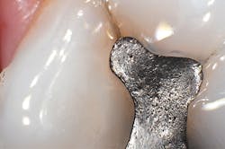
Intraoral photographs are imperative in my diagnostic process. To show a patient a lesion with a mirror on an upper-left second molar is nearly impossible. Viewing that area magnified 20 times on a 32-inch screen is a much more explanatory visual aid. I use the high-definition DEXcam 4 HD (DEXIS) to take my intraoral photos. These images are useful in multiple ways—for example, as an education tool for patients and a reference point for lesions on the tissue (so we can look back to see progression at future appointments). If patients don’t want to address an issue immediately, we can show how their problems, such as caries or broken fillings, have gotten progressively worse. Then they can be confident that it’s time to act.
Intraoral photos are also helpful in conjunction with radiographs for insurance claims that can be filed electronically. An old mesio-occlusal or L-shaped restoration can all look tiny on a radiograph. But when you see them on intraoral photographs that show they extend 80% of the width of the occlusal table of the tooth buccolingually, the insurance company can clearly see that the tooth is not restorable with a traditional filling material but will need an onlay or a crown. Another use for intraoral photos is communication with the lab. I am able to take a picture of a crown in place and send it to the lab with an explanation to correct shape issues.
DEXcam intraoral photo
To get a true picture of the mouth, digital x-rays, caries detection devices, and intraoral photographs work hand in hand. If you can have multiple sources of diagnostic reference available, why would you say no to using them?
I am focused on doing what’s best for my patients, but I can’t help them if they don’t understand their problems or realize that I am seeking the best solution. They have to want treatment. It is so satisfying when they see and finally understand what they need for appropriate dental care. I don’t know if these imaging modalities set my practice apart from others; I just believe it’s a standard of care. There are only advantages to having these tools at my fingertips.
Gary Tomack, DDS, FAGD, attended Howard University College of Dentistry and completed residency at Harlem Hospital–Columbia University, where he became an instructor and then chief of cosmetic dentistry—a position he held for 28 years. He has been in practice in the Murray Hill area of Manhattan since 1983 and lectures at local study clubs and at Harlem Hospital–Columbia University. Dr. Tomack has no financial interests in DEXIS LLC or KaVo Kerr.


