Guide to going digital: Intermediate level
Part TWO of a three-part series
Edward Shellard, DMD
You don't know what you don't know. This statement applies to various aspects of dentistry, but nowhere is it more applicable than when it comes to diagnostics. Imaging plays an important role in providing you with all the information you need to diagnose a case and communicate effectively with the patient.
In this second part of my three-article series, I would like to explore cone beam computed tomography (CBCT), as well as the various ways it can help the way you practice. From allowing you to make more confident and accurate diagnoses to improving patient education and case acceptance, 3-D imaging has changed the field of dentistry for the better.
More information for diagnosis
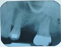
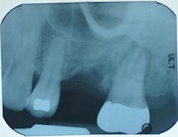
A root fracture, perforated canal, and periapical lesion are all common problems that can appear normal on a 2-D radiograph. Without CBCT imaging, you don't have all of the information necessary to diagnose the issue as superimposition and magnification can make it difficult to see every part of the anatomical structure.
In the past, diagnosing a root fracture was difficult. Based on the patient's description of the pain and a radiograph, a dentist may have prepared the tooth for a crown to see how it felt. When that didn't address the pain, a root canal would have been recommended. After that, the only option may have been extraction. This process was frustrating not only to the patient-who spent time and money to get the pain relieved-but also to the doctor who wanted to address the problem.
Today, we can eliminate this clinical progression by having all of the information upfront. This results in better treatment outcomes and patient care.
2-D periapical image
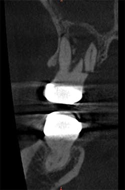
3-D imaging
Root fracture as seen on an axial scan view. Clinical photos courtesy Dr. Bradley McAllister, DDS, PhD
Improved patient communication
When using 2-D radiographs alone, you can't fully tell the patient the whole situation; with a 3-D volume, on the other hand, you have the extra information necessary so you can share more options with your patients. This gives patients the tools necessary to make informed decisions about their own care and increases your case acceptance rate.
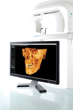
Not only that, but patients are impressed when they can see their own skull on the monitor. This enables you to point out any problem areas in a way that makes sense to the patient as well as impress them with your cutting-edge technology.
Factors to consider when implementing CBCT
Many doctors are worried about the costs associated with purchasing a CBCT system. One thing I would ask you to consider is the number of patients you are referring to a third-party imaging center for scans. By keeping patients within your office, you not only have the opportunity to provide the scan as a service, but it's more likely that the patient will actually have it done and accept the treatment plan.
Another factor to consider is space. While CBCT units of the past were quite large, there are now compact units that don't take up as much practice real estate. And, since they are capable of capturing both panoramic and CBCT images, you benefit from different modalities all in one machine.
The takeaway
With 2-D imaging alone, you're only getting a small piece of the puzzle. Incorporating CBCT imaging into your practice will not only change the way you diagnose and plan treatments, but also how you communicate with your patients. All in all, 3-D imaging allows you to know more-and reduces what you don't know.
Edward Shellard, DMD, has more than two decades of clinical and executive experience in the dental industry. Since joining Carestream Dental in 2008, Dr. Shellard has driven global marketing strategy and product development efforts for the latest innovations in dental and practice-management technology.


