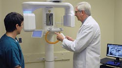
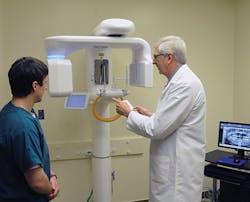
David Gane, DDS, interviews
University of Tennessee Professor Jeffrey Brooks, DMD
Cone Beam Computed Tomography is becoming the standard of care for improved diagnosis and treatment planning across the dental industry. Part of this trend is the adoption of advanced technologies by new dental school graduates looking to provide a higher standard of care and additional treatment options as they begin their dental careers. To find out how today's new dental professionals are learning about CBCT imaging and other innovations, I spoke with my colleague, Dr. Jeffrey Brooks, an associate professor in the Department of Oral and Maxillofacial Surgery at the University of Tennessee Health Science Center College of Dentistry.
Dr. Gane: Dr. Brooks, you're currently teaching at the University of Tennessee (UT), but you also graduated from the school's College of Dentistry. This puts you in a unique position to provide insight regarding how education in our industry has changed over the years. How are today's students learning about the latest imaging technology?
Dr. Brooks: As recently as five years ago, most dental schools weren't teaching undergraduate or even post-graduate students to use technology such as CBCT imaging. The lack of exposure to this technology was multi-factorial. Most dental schools in the U.S. didn't have these systems in their facilities, and the faculty were not qualified to train the students on how to utilize this advanced imaging technology to better diagnose and treat their patients. Primarily, this was because they didn't have access to this technology during their dental school training. Traditionally it was left to the manufacturers of imaging systems to provide the main source of education and information on advanced imaging technology via extensive continuing education (CE) courses outside the academic environment.
Now, educational institutions are introducing both undergraduate and post-graduate students to these advanced imaging modalities at the earliest stages of their education. At UT, we've implemented a didactic curriculum where experienced faculty members train students on how and when they should utilize traditional 2-D imaging modalities versus 3-D CBCT imaging. Students at the UTHSC College of Dentistry have access to the didactic elements of 3-D CBCT imaging in their classes and hands-on experience with these units in the various specialty departments within the school. They are able to obtain 3-D scans for better diagnostic imaging, which results in better overall treatment of the patients at the school.
Dr. Gane: What resources does UT have to support this curriculum?
Dr. Brooks: The dental school has eight extraoral imaging systems available to dental students, which include panoramic, cephalometric, and CBCT units. Seven of the eight systems have 3-D imaging capabilities, which I would guess is more than any other dental school in the nation. However, mentorship is one of the keys to an effective learning experience, which is why we have qualified faculty members sit down on a one-to-one basis with students to show them how to use the imaging systems in conjunction with the feature-rich software.
Dr. Gane:What about post-graduation? If a student plans to run his or her own practice, how do you prepare him or her to make informed decisions about purchasing imaging technologies?
Dr. Brooks: We provide access to products from various manufacturers, which allows the students to see for themselves which systems offer the best image quality, ease of use, and lowest radiation dose. The students often migrate to the system they enjoy the most. In the Oral and Maxillofacial Surgery department (OMS), we are using Samsung's Rayscan Alpha - Expert 3-D digital imaging system. Students from outside the OMS program come to our department to use the system due to its image quality and ease of use.
Like the rest of the faculty, I personally take time with our students at the Rayscan Alpha to educate them on the system's panoramic and CBCT modalities, as well as how to properly manipulate radiographs in the imaging software. As an example, a student may have a patient's traditional panoramic image that includes a questionable lesion in the mandible that can't be identified as an artifact or part of the pathology, so he or she will capture additional panoramic and CBCT images to obtain more clinical information.
Dr. Gane:Beyond an increased knowledge of CBCT technology and its clinical applications, is there anything else that students are learning from using the latest imaging technology?
Dr. Brooks: There are certainly several intangibles that come with being familiar with the latest imaging technology. Three-dimensional images simplify diagnosis, which in turn improves treatment planning and decreases patient morbidity. In the end, you have a happy practitioner and a happy patient. Students are able to graduate and be more productive immediately because they already have this technological familiarity and knowledge. Additionally, the time savings and diagnostic advantages afforded by the technology improve patient relationships and build trust with referring dentists. By helping to develop the technological skill sets of the next generation of clinicians, we leave them better prepared to provide superior care and grow their practices into successful businesses.
Dr. Gane:Not every practice has an imaging unit with CBCT capabilities. How do these students react when they join a practice after graduation that doesn't have this technology?
Dr. Brooks: A digital workflow with 3-D imaging capabilities is definitely what our graduates are accustomed to when they start their careers; in fact, it becomes an expectation that this technology will be available when they join a practice. When they transition into a practice that isn't utilizing digital imaging or doesn't have access to advanced CBCT technology, their appreciation for the diagnostic and workflow advantages of the technology often makes them an advocate for upgrading the practice. These new graduates recognize the clinical and economic advantages of having CBCT technology available.
Dr. Gane: It sounds like there's a great program in place at UT to prepare tomorrow's practitioners. But on the topic of upgrading practices, what about today's dentists who did not have the benefit of learning about 3-D imaging in school? What recommendations would you offer them to get some hands-on experience with the latest technology?
Dr. Brooks: There are a lot of great CE programs available that cover the latest innovations, including developments in CBCT and CAD/CAM technology. There are courses tailored to all levels of familiarity with the subject matter, whether it's in an introductory setting or a more advanced environment. In fact, UT has plans to offer CE programs on 3-D imaging in the near future. Increasing the diagnostic capabilities of a practice with advanced imaging technology opens the door to a whole new world of treatment options that can positively impact a practice for years to come. If anyone has specific questions, they are welcome to contact me directly at [email protected] - I'm always happy to help!
David Gane, DDS, is the president and CEO of LED Medical Diagnostics. Dr. Gane is a member of the Canadian Dental Association, the American Dental Association, and the American Academy of Oral and Maxillofacial Radiology, where he served as Chairman of the Corporate Liaison Committee.
Jeffrey Brooks, DMD, is a board certified Oral and Maxillofacial Surgeon. He maintains a position as associate professor in the Department of Oral and Maxillofacial Surgery as well as serving as its director of 3-D Imaging. Dr. Brooks currently serves as Vice President of Imaging for LED Dental.


