I love going to dental conferences, and I love to learn new things that help me get better at my craft. Give me great locations and great speakers, and I’m all in. I enjoy meeting other clinicians and hearing about what they’re doing in their practices. Sometimes that’s the best CE I get! But it never fails that I hear stress in many of their voices, and I can see it on their faces. I used to be that guy.
We all know it’s hard to run a successful dental practice—overhead goes up and we deal with rising costs, debt, inflation, demanding patients, and challenges with staff. Being a high-end, successful dentist and business owner comes with major challenges.
I learned long ago that you can’t cut costs on your way to prosperity. I also learned that you must invest in your practice and yourself. When I decided to integrate dental implants into my practice, I made a big investment in products, equipment, and myself. I took tons of training because I wanted to make sure I did everything right for me and my patients.
It was the same when I bought a CEREC, clear aligners, CBCT, and other new technologies. It wasn’t cheap or quick, but the payoff in my patient care, job satisfaction, and finances has been well worth it.
You may also be interested in ... How AI can (and will) impact your dental practice
Over the years, as I built my practice and honed my skills, I decided that if I could find technologies that would make my practice and me better, I would make the investment no matter the cost or time commitment.
As I continue to explore new technologies, I ask if they improve my practice, patient care, job enjoyment, and help me make more money. Money isn’t the most important thing, but it’s important. I’ve passed these principles along to my wife (who practices with me) and my son (who has opened his own private practice nearby).
A new technology called 3D tomosynthesis
I recently heard about a new technology—3D tomosynthesis—from a friend. I was an early adopter of CBCT, so I was familiar with 3D tomography. I’ve always appreciated what 3D provides me with cone beam, so I was compelled to dig deeper. The product is the Portray System from Surround Medical Systems Inc. The system looks like the typical wall-mounted 2D units in my practice (figure 1).
More data any way you slice it
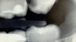
What’s unique about this system is that it uses nanotube technology to capture standard 2D images (figure 2) and gives you the option of taking a tomosynthesis image, which captures multiple images from several angles (figure 3). Images are compiled into a sliced volume and dissected into 0.5 mm or 0.1 mm slices that you can scroll through, rotate, enlarge, measure, and adjust. The system can “un-overlap” many teeth; this is convenient because with regular 2D x-rays, I had to retake the image at different angles and often with little success.
As I’ve grown more comfortable with the data I get from Portray, my level of satisfaction with patient interactions has increased because I can show my patients things even I couldn’t see before. Patients can tell when you’re excited about finding something new. Even though I’ve only had the system for about a year, I’ve already had a nice return on investment. By my best estimate, I’d say the system paid for itself within about six to eight months.
I see more complications and pathologies when using 3D tomosynthesis than I ever did with my old 2D images (figure 4). I can treat more because I can see more bone loss, interproximal caries, fractures, resorptions, abscesses, and more (figure 5).
In fact, twice today I was able to see something with Portray that wasn’t visible in the original 2D x-ray (figure 6). I don’t always take 2D images now, but I do when I think the case is straightforward or the patient has very little dental history. I often end up taking a tomosynthesis image just to be safe. With the 3D image I can scroll from buccal to lingual through the tooth and get much more data (figure 7). The new system has replaced my 2D PAs and bitewings.
Where did 3D tomosynthesis come from?
3D tomosynthesis is new to dentistry, but it’s not new to the medical field. Lifting a page from the mammography industry’s playbook, 3D tomosynthesis has been a decades-old game changer. 2D imaging was once the staple for radiologists and their imaging, just like it is in dentistry today. Now, 3D tomosynthesis is the gold standard in mammographic imaging and can be used to detect over 40% more cancers than 2D imaging can. Most radiologists have moved away from 2D, and now over 90% of all mammographic images are taken with 3D tomosynthesis.
Fortunately, 3D intraoral tomosynthesis found its way into the dental field. Based on the findings from a study out of the University of North Carolina, dentists who use 3D tomosynthesis for intraoral x-rays can detect approximately 36% more caries (figures 8 and 9).1 That’s tremendous!
Is 3D intraoral tomosynthesis a headache to use?
As with any new technology, I’m always concerned with ease of integration. I believe this is one of the main benefits of Portray. It integrates seamlessly into the practice; the software is plug-and-play with virtually no learning curve. The workflow is the same as with stationary 2D intraoral imaging. Using Portray has been a real no-brainer for my practice. The image capture process is easy for my team because it’s compatible with many of the top practice management systems. We get more data with this new software, which allows us to be more precise in our patient care.
What about radiation?
Incredibly, the radiation levels for 3D tomosynthesis are comparable to 2D x-ray systems—and much lower than a cone beam. While it doesn’t take the place of a CBCT unit, 3D tomosynthesis will bridge the gap between your 2D and cone beam. It does this by providing high-resolution sliced images of a focused area. The more information I can get on the front end, the quicker I can diagnose a case and not disrupt the workflow by having to move the patient to the CBCT.
More about radiation ... How much radiation is in a dental x-ray? You might be surprised
This technology has been great for me, my team, our patients, and the practice. It’s yet another area where improved technology makes my life easier. I believe I’m a better clinician now, my revenue has increased, and I have less stress.
The future of dental imaging awaits
Embrace the future of dental imaging and embark on a journey that’s changing the face of dentistry. The Portray System isn’t just a tool; it’s a statement of your commitment to excellence. It’s time to enhance your patient care, unleash your potential, and discover a realm of possibilities once hidden. Now, innovation can meet aspiration.
Editor's note: This article appeared in the November 2023 print edition of Dental Economics magazine. Dentists in North America are eligible for a complimentary print subscription. Sign up here.
Reference
1. Mol A, Platin E, Gaalaas LR. Comparison of a stationary intraoral tomosynthesis using a carbon nanotube field emission x-ray source and conventional two-dimensional imaging for proximal caries detection. An observer study. The University of North Carolina at Chapel Hill.
David C. Koski, DMD, a graduate of Case Western Reserve University School of Dentistry, has a particular expertise in cosmetic dentistry and implantology. He is the president of the Koski DePaul Dental Group and CEO of Jupiter BioLife. He is a member of the American Academy of Implant Dentistry and a provider for the Center of Advanced Rejuvenation and Esthetics. In addition to his work in private practice, Dr. Koski is a facial esthetics educator at Advanced PRF (Platelet-Rich Fibrin) Education.


