MAXIMIZING the RETUNRN ON INVESTMENT of an OPERATING MICROSCOPE

by David Clark, DDS
When I was approached to write this article, I scratched my head at first. Dentists buy microscopes out of a passion for excellence. Profit is often an afterthought. Yet, the creation of a microscope-centered practice can yield a rich return on investment (ROI) regardless of the type of practice.
Successful companies focus simultaneously on two factors that influence the bottom line: reducing expenses and increasing revenues. These two factors also can be thought of as offense (income) and defense (reducing costs). Throughout the article we will highlight both parameters.
The following two examples embody the financial implications of the operating microscope. We will explore case 1, the $10,000 molar endo, and case 2, the $35 sealant.
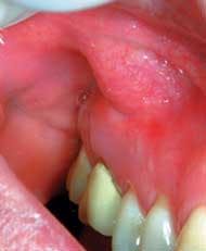
The $10,000 endo
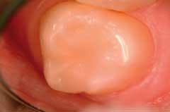
When a family quietly leaves a practice because of microscopic failures to their expensive treatments, the real costs to the dentist are substantial. Figure 1 shows low magnification of a patient who left his previous restorative dentist because of failing endodontic therapy. He has had periodic swelling for years. The previous dentist, who does not own a microscope, missed the MB2 canal system. The patient and his family of four had seen this dentist for years. Do the math: The family of four came in for exams, X-rays, and cleanings for 10 years. Total future revenues lost will exceed $10,000. Bad offense!
The $35 sealant
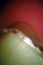
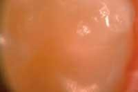
Once I began to do examinations with the microscope, I was embarrassed by the poor condition of many of the sealants that we had worked so carefully to place. Studies have shown the 10-year failure rate of traditional sealants is as much as 90 percent (see figure 3, 4). This can result in multiple re-treatments — at no charge or reduced fees, of course. Bad defense!
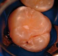
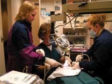
We now perform a very different procedure under the operating microscope (See figure 5,6.).
The miracle of the microscope
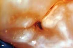
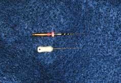
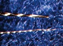
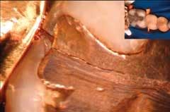
Low magnification of these endodontic files (figure 7) is unimpressive. But at 24X (figure 8), we see the metal is unwinding. One more insertion would have resulted in instrument separation, costing the practitioner minutes or hours to address. When the patient learns about the mishap, future relations are strained or broken. This type of incident accounts for a significant portion of dental malpractice lawsuits. Bad offense and bad defense!
When clinicians use an operating microscope throughout an endodontic procedure, they will likely see problems before disaster strikes.
The creation of a microscope-centered practice
The single most important element for realizing a significant ROI and creating a microscope-centered practice is to allow patients and staff to see the same incredible view the doctor sees. Show your patients a live or recorded video broadcast — or both! — of their mouths as you examine and treat them. Using a live, microscope video feed is superior to a pencil- sized intraoral camera for two reasons: Microscope video is hands-free and interruption free. Because of the unique camera mounting on the microscope, broadcasting the images becomes a non-event. Broadcasting live treatment as it progresses has never been easily accomplished until the advent of the microscope. Patients are mesmerized and grateful for the doctor's commitment to precision.
As I talk to dentists from around the world who own microscopes, I am continually amazed that so many do not use the power of video to demonstrate the value of precision dentistry and elegant diagnosis.
The most common problem is the misconception that the doctor needs a complex technical system for this documentation. You don't! Here is a simple, low-tech plan to position your practice as "microscope centered:"

• Make sure you have a video camera installed when you purchase your microscope. If you own a microscope without a video feed, install one now!
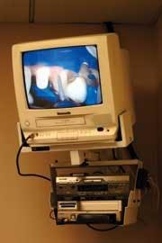
• An inexpensive television or LCD screen should be placed in patient's view and should always be turned on (See figure 9).
• The key to a microscope-centered practice is taking microvideo and or microphotography of the tooth and soft-tissue portion of the comprehensive new-patient exam.(See figure 10). Mini digital video is the best format and stores easily in the patient chart.
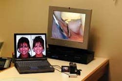
• Present the video later as part of the comprehensive treatment consult. (See figure 11.) The one exception to live-time microscopic video broadcast is the new-patient exam. I find it distracting and it lessens the presentation value.
This simple process requires almost no technical learning curve and no interruption of your clinical routine. It will literally transform your practice by revolutionizing the most important appointment — the new patient experience.
Clinicians who convert to this microscope-centered concept report finding a wealth of subtle disease that they could not see before:
• Micro pathology — This new term describes important pathology that is either invisible or not compelling at less than 12X. This includes signs of occlusal disease (telltale facets and enamel loss); early incomplete fractures; micro leakage; early recurrent decay; and isolated periodontal inflammation surrounding crude dentistry that violates the three new parameters of marginal integrity.

• Dramatic results —Patient motivation is, in a word, incredible. Average adults over 30 present with two posterior teeth with fairly dramatic, early incomplete coronal fractures that their last dentist never saw. Many of these teeth have a history of symptoms. In the average practice, it will take several years of several procedures per week to treat these teeth.
• Acceptance — Patients begin to understand and accept the value of quadrant and comprehensive dentistry instead of tooth-at-a-time dentistry.
A patient's point of view
Patients understand and value the impact of an operating microscope on their dental health. Jim, who has been with my practice since I first began (more than 17 years ago) says, "I believe that the operating microscope has enabled Dr. Clark to offer a level of restorative dentistry unheard of only a few years ago. Dr. Clark and his staff offer a level of care that is on the cutting edge of dental science."
Jim has referred more than 20 individuals who have become full-time patients in my practice; they, in turn have referred a substantial number of their own friends and family. I asked this same gentleman to explain what it's like to watch live treatment on the television while I use the microscope. He says, "I can see for myself the true precision and craftsmanship of Dr. Clark's work.
"If something were to happen to Dr. Clark, or if he should retire, I would never seek care from a dentist who did not use a microscope. It is impossible to receive the level of care that I have come to expect without it."
Pat has this to say about the impact of microscope use on her oral health: "The operating microscope is amazing. I have become accustomed to seeing for myself a whole new world. The precision of Dr. Clark's diagnoses and clinical work is truly impressive.
"The microscope made it possible for him to determine which of my teeth was really cracked after I fell and hit the pavement with my jaw. The tooth with the subtler crack was the problematic one. I am grateful to Dr. Clark and the microscope for the correct diagnosis."
Anatomy of a bankrupt practice
We have had the unique experience of absorbing the patients and partial staff of a bankrupt dental practice. The following factors contributed to this practice's demise; all of them could be reduced or eliminated by incorporating an operating microscope into the practice.
! Multiple re-do's
! Insurance-dependent patients
! The $35 sealant
! Patients refusing treatment because previous treatments shred floss, stains, are micro-leaking and uncomfortable, or have required repair or re-treatment
! Patient and doctor could not see the micro pathology (structurally unsound teeth, micro leakage and early recurrent decay)
! Constant interruptions from toothaches and broken teeth
Activity vs. productivity
A traditional dental practice is like a beehive. Patients come and go, operatories and instruments are set up, broken down, and set up again. In the aforementioned bankrupt practice, there was substantial activity. The problem with feeling busy is that it can lull the team into a false sense of success. In a microscope-centered practice, a focus on productivity instead of activity is a natural by-product.
Good for your practice � Good for you
Many of my colleagues are slowing down or retiring prematurely because of chronic, posture-related health problems. Do the math: 10 (years) x $150,000 (average annual salary) = $1.5 million lost income. Cataclysmically bad offense! Using the microscope creates ideal posture. Clinicians report an immediate reduction in back, shoulder, and neck problems. There are many clinicians who have switched to the microscope because of chronic back, shoulder, and neck pain; they report a dramatic improvement in comfort. It is more prudent, however, to initiate good posture before career ending symptoms develop. Using an operating microscope has extended the careers of many neurosurgeons and dentists. The operating microscope repairs sore backs, tired eyes, and worn-out attitudes.

Other intangible benefits to using a microscope are:
• Confidence
• Pride
• Peace of mind
• Eliminating the "iffy" diagnosis
These benefits are as important as any financial windfall.
Microscopic precision and visualization often eliminates the emotional and financial toll of failures that could have been avoided. Whenever disgruntled patients leave the practice, it takes the wind out of the office's emotional sails. An operating microscope can help practices avoid the letter no one wants to see, such as the following actual letter:
"Dear Dr. J.: We have been contacted by your former patient, and here are our findings: Open margins ... the patient is unhappy ... Four veneers will need to be redone at a cost of $7,500 ... please send check."
Continuing education in microscope dentistry
Owning a microscope without continuing education is like buying a car with no seats. Consequently, many doctors have "parked" their microscopes in the corner with infrequent use because they feel uncomfortable using them. The first wave of dentists to use microscopes were self-taught and let me tell you — we learned slowly. Today, there are excellent courses, but you need to know where to find them. The Academy of Microscope Enhanced Dentistry provides the broadest array of lectures and hands-on courses. Contact information is at www.microscopedentistry.com. The Newport Coast Oral Facial Institute provides microscope-centered teaching at ncofi.org. A final source is Precision Aesthetics Northwest. Information is available at lifetimedentistry.net.
Positioning your practice for the future
High-volume dentistry is one way to generate significant revenues, as the coming wave of franchise dentistry indicates. In these types of practices, the microscope may collect dust. On the other hand, there are many clinicians who want to provide more comprehensive dentistry, spend more time at the new-patient examination, and improve the quality of the treatment they provide. These clinicians deserve to make good livings but often have difficulty with the bottom line when they begin to overhaul their practices.
The operating microscope combined with constant visual feedback to patients is the key to providing unparalleled excellence and attaching a tangible value and product differentiation. Today's consumer follows the old Chinese saying, "What I hear, I forget. What I see, I remember." Telling your patient how careful your margins are has little impact. Delivering on that promise and showing them is everything.
A dentist who attended one of our courses tracked me down later that day on the busy convention floor. He said, "David, I just want you to know that I was moved by the experience, and I just bought my first microscope. It will change my practice; I think it may even change my life."
It did. And it will!.